- PDB-6jl3: Crystal Structure of the UBL domain of Plasmodium Falciparum Dsk2 -
+
Open data
ID or keywords:
Loading...
-
Basic information
Entry
Database: PDB / ID: 6jl3
Title
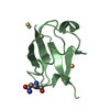
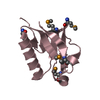
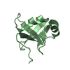
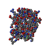
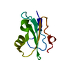
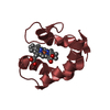
Crystal Structure of the UBL domain of Plasmodium Falciparum Dsk2
Components
Ubiquitin domain-containing protein DSK2,putative
Keywords
SIGNALING PROTEIN / Ubiquitin binding / shuttle factor / ubiquitin
Function / homology
Function and homology information
Cargo recognition for clathrin-mediated endocytosis / polyubiquitin modification-dependent protein binding / ubiquitin-dependent protein catabolic process / cytosol Similarity search - Function
Mass: 8672.269 Da / Num. of mol.: 1 / Fragment: UNP residues 1-77 Source method: isolated from a genetically manipulated source Details: The initial two amino acid (MA) and the last three amino acid (AMA) electron density were missing. Source: (gene. exp.) Plasmodium falciparum (malaria parasite P. falciparum) Gene: PF3D7_1113400 / Plasmid: pETM11 / Details (production host): His-tag / Cell line (production host): DE3 / Production host: Escherichia coli B83 (bacteria) / Strain (production host): B83 / References: UniProt: Q8IIM8
In the structure databanks used in Yorodumi, some data are registered as the other names, "COVID-19 virus" and "2019-nCoV". Here are the details of the virus and the list of structure data.
Jan 31, 2019. EMDB accession codes are about to change! (news from PDBe EMDB page)
EMDB accession codes are about to change! (news from PDBe EMDB page)
The allocation of 4 digits for EMDB accession codes will soon come to an end. Whilst these codes will remain in use, new EMDB accession codes will include an additional digit and will expand incrementally as the available range of codes is exhausted. The current 4-digit format prefixed with “EMD-” (i.e. EMD-XXXX) will advance to a 5-digit format (i.e. EMD-XXXXX), and so on. It is currently estimated that the 4-digit codes will be depleted around Spring 2019, at which point the 5-digit format will come into force.
The EM Navigator/Yorodumi systems omit the EMD- prefix.
Related info.:Q: What is EMD? / ID/Accession-code notation in Yorodumi/EM Navigator
Yorodumi is a browser for structure data from EMDB, PDB, SASBDB, etc.
This page is also the successor to EM Navigator detail page, and also detail information page/front-end page for Omokage search.
The word "yorodu" (or yorozu) is an old Japanese word meaning "ten thousand". "mi" (miru) is to see.
Related info.:EMDB / PDB / SASBDB / Comparison of 3 databanks / Yorodumi Search / Aug 31, 2016. New EM Navigator & Yorodumi / Yorodumi Papers / Jmol/JSmol / Function and homology information / Changes in new EM Navigator and Yorodumi
 Movie
Movie Controller

 Yorodumi
 Open data
 Basic information
 Components
 Keywords
 Function and homology information
 X-RAY DIFFRACTION /
  Authors
 India, 1items
  Citation
 Structure visualization
 Downloads & links
 Other downloads


 PDBj
PDBj

 Assembly

 Mass: 18.015 Da / Num. of mol.: 82 / Source method: isolated from a natural source / Formula: H2O
 Sample preparation
 / Beamline: ID23-1 / Wavelength: 1 Å
 Processing