 Movie
Movie Controller


+ Open data

- Basic information
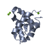
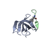
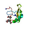
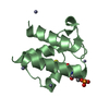
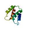
| Entry | Database: PDB / ID: 3bqq | ||||||
|---|---|---|---|---|---|---|---|
| Title | Crystal Structure of Human Saposin D (triclinic) | ||||||
 Components Components | PROACTIVATOR POLYPEPTIDE | ||||||
 Keywords Keywords | LIPID BINDING PROTEIN / SAPOSIN / SPHINGOLIPID ACTIVATOR PROTEIN / ACID CERAMIDASE / FARBER DISEASE / LIPID METABOLISM / LYSOSOME / SPHINGOLIPID METABOLISM | ||||||
| Function / homology |  Function and homology information Function and homology informationganglioside GM1 transport to membrane / ganglioside GM2 binding / ganglioside GM3 binding / ganglioside GP1c binding / ganglioside GM1 binding / ganglioside GT1b binding / epithelial cell differentiation involved in prostate gland development / prostate gland growth / sphingolipid metabolic process / Glycosphingolipid catabolism ...ganglioside GM1 transport to membrane / ganglioside GM2 binding / ganglioside GM3 binding / ganglioside GP1c binding / ganglioside GM1 binding / ganglioside GT1b binding / epithelial cell differentiation involved in prostate gland development / prostate gland growth / sphingolipid metabolic process / Glycosphingolipid catabolism / lysosomal transport / azurophil granule membrane / regulation of lipid metabolic process / lysosomal lumen / regulation of autophagy / Peptide ligand-binding receptors / phospholipid binding / enzyme activator activity / adenylate cyclase-inhibiting G protein-coupled receptor signaling pathway / Platelet degranulation / late endosome / protease binding / gene expression / scaffold protein binding / G alpha (i) signalling events / lysosome / lysosomal membrane / Neutrophil degranulation / protein homodimerization activity / : / extracellular exosome / extracellular region / identical protein binding / plasma membrane Similarity search - Function | ||||||
| Biological species |  Homo sapiens (human) Homo sapiens (human) | ||||||
| Method |  X-RAY DIFFRACTION / MOLECULAR REPLACEMENT / Resolution: 2 Å X-RAY DIFFRACTION / MOLECULAR REPLACEMENT / Resolution: 2 Å | ||||||
 Authors Authors | Prive, G.G. / Popovic, K. | ||||||
 Citation Citation | Journal: Acta Crystallogr.,Sect.D / Year: 2008 Title: Structures of the human ceramide activator protein saposin D. Authors: Popovic, K. / Prive, G.G. | ||||||
| History |
|
- Structure visualization
| Structure viewer | Molecule: MolmilJmol/JSmol |
|---|
- Downloads & links
-Download
| PDBx/mmCIF format | 3bqq.cif.gz | 77.7 KB | Display | PDBx/mmCIF format |
|---|---|---|---|---|
| PDB format | pdb3bqq.ent.gz | 59.6 KB | Display | PDB format |
| PDBx/mmJSON format | 3bqq.json.gz | Tree view | PDBx/mmJSON format | |
| Others |  Other downloads Other downloads |
-Validation report
| Arichive directory | https://data.pdbj.org/pub/pdb/validation_reports/bq/3bqqftp://data.pdbj.org/pub/pdb/validation_reports/bq/3bqq | HTTPS FTP |
|---|
-Related structure data


-Links
 PDBj
PDBj


- Assembly
| Deposited unit | 
| ||||||||
|---|---|---|---|---|---|---|---|---|---|
| 1 | 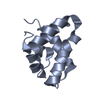
| ||||||||
| 2 | 
| ||||||||
| 3 | 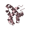
| ||||||||
| 4 | 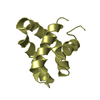
| ||||||||
| Unit cell |
|
-Components
| #1: Protein | Mass: 8926.332 Da / Num. of mol.: 4 / Fragment: SAPOSIN D, RESIDUES 405-484 Source method: isolated from a genetically manipulated source Source: (gene. exp.) Homo sapiens (human) / Gene: PSAP / Plasmid: PET-16 / Production host:  #2: Water | ChemComp-HOH / |  Mass: 18.015 Da / Num. of mol.: 314 / Source method: isolated from a natural source / Formula: H2O Mass: 18.015 Da / Num. of mol.: 314 / Source method: isolated from a natural source / Formula: H2OHas protein modification | Y | |
|---|
-Experimental details
-Experiment
| Experiment | Method: X-RAY DIFFRACTION / Number of used crystals: 1 |
|---|
- Sample preparation
| Crystal | Density Matthews: 2.84 Å3/Da / Density % sol: 56.75 % |
|---|---|
| Crystal grow | Temperature: 298 K / Method: vapor diffusion, hanging drop / pH: 4.6 Details: PEG 4K, ZINC SULFATE, SODIUM ACETATE BUFFER, PH 4.6, VAPOR DIFFUSION, HANGING DROP, temperature 298.0K |
-Data collection
| Diffraction | Mean temperature: 100 K |
|---|---|
| Diffraction source | Source: ROTATING ANODE / Type: BRUKER AXS MICROSTAR / Wavelength: 1.5418 / Wavelength: 1.5418 Å |
| Detector | Type: BRUKER SMART 6000 / Detector: CCD / Date: Mar 24, 2007 |
| Radiation | Protocol: SINGLE WAVELENGTH / Monochromatic (M) / Laue (L): M / Scattering type: x-ray |
| Radiation wavelength | Wavelength: 1.5418 Å / Relative weight: 1 |
| Reflection | Resolution: 2→65 Å / Num. obs: 24429 / % possible obs: 92.6 % / Redundancy: 3.52 % / Rmerge(I) obs: 0.056 / Net I/σ(I): 20.4 |
| Reflection shell | Resolution: 2→2.05 Å / Redundancy: 1.4 % / Rmerge(I) obs: 0.1406 / Mean I/σ(I) obs: 6.73 / % possible all: 91.2 |
- Processing
| Software |
| ||||||||||||||||||||||||||||||||||||||||||||||||||||||||||||||||||||||||||||||||||||||||||
|---|---|---|---|---|---|---|---|---|---|---|---|---|---|---|---|---|---|---|---|---|---|---|---|---|---|---|---|---|---|---|---|---|---|---|---|---|---|---|---|---|---|---|---|---|---|---|---|---|---|---|---|---|---|---|---|---|---|---|---|---|---|---|---|---|---|---|---|---|---|---|---|---|---|---|---|---|---|---|---|---|---|---|---|---|---|---|---|---|---|---|---|
| Refinement | Method to determine structure: MOLECULAR REPLACEMENT Starting model: SAPOSIN D P212121 Resolution: 2→64.55 Å / Cor.coef. Fo:Fc: 0.941 / Cor.coef. Fo:Fc free: 0.92 / SU B: 3.71 / SU ML: 0.106 / Cross valid method: THROUGHOUT / ESU R: 0.185 / ESU R Free: 0.169 / Stereochemistry target values: MAXIMUM LIKELIHOOD / Details: HYDROGENS HAVE BEEN ADDED IN THE RIDING POSITIONS
| ||||||||||||||||||||||||||||||||||||||||||||||||||||||||||||||||||||||||||||||||||||||||||
| Solvent computation | Ion probe radii: 0.8 Å / Shrinkage radii: 0.8 Å / VDW probe radii: 1.4 Å / Solvent model: MASK | ||||||||||||||||||||||||||||||||||||||||||||||||||||||||||||||||||||||||||||||||||||||||||
| Displacement parameters | Biso mean: 21.496 Å2
| ||||||||||||||||||||||||||||||||||||||||||||||||||||||||||||||||||||||||||||||||||||||||||
| Refinement step | Cycle: LAST / Resolution: 2→64.55 Å
| ||||||||||||||||||||||||||||||||||||||||||||||||||||||||||||||||||||||||||||||||||||||||||
| Refine LS restraints |
| ||||||||||||||||||||||||||||||||||||||||||||||||||||||||||||||||||||||||||||||||||||||||||
| LS refinement shell | Resolution: 2→2.052 Å / Total num. of bins used: 20
|
