 Movie
Movie Controller


+ Open data

- Basic information
| Entry | Database: PDB / ID: 3lef | ||||||
|---|---|---|---|---|---|---|---|
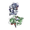
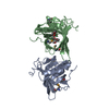
| Title | Crystal structure of HIV epitope-scaffold 4E10_S0_1Z6NA_001 | ||||||
 Components Components | Uncharacterized protein 4E10_S0_1Z6NA_001 (T18) | ||||||
 Keywords Keywords | IMMUNE SYSTEM / EPITOPE-SCAFFOLD | ||||||
| Function / homology | Glutaredoxin / Glutaredoxin / 3-Layer(aba) Sandwich / Alpha Beta Function and homology information Function and homology information | ||||||
| Biological species | ARTIFICIAL GENE (others) | ||||||
| Method |  X-RAY DIFFRACTION / MOLECULAR REPLACEMENT / Resolution: 2.3 Å X-RAY DIFFRACTION / MOLECULAR REPLACEMENT / Resolution: 2.3 Å | ||||||
 Authors Authors | Holmes, M.A. | ||||||
 Citation Citation | Journal: Structure / Year: 2010 Title: Computational Design of Epitope-Scaffolds Allows Induction of Antibodies Specific for a Poorly Immunogenic HIV Vaccine Epitope. Authors: Correia, B.E. / Ban, Y.E. / Holmes, M.A. / Xu, H. / Ellingson, K. / Kraft, Z. / Carrico, C. / Boni, E. / Sather, D.N. / Zenobia, C. / Burke, K.Y. / Bradley-Hewitt, T. / Bruhn-Johannsen, J.F. ...Authors: Correia, B.E. / Ban, Y.E. / Holmes, M.A. / Xu, H. / Ellingson, K. / Kraft, Z. / Carrico, C. / Boni, E. / Sather, D.N. / Zenobia, C. / Burke, K.Y. / Bradley-Hewitt, T. / Bruhn-Johannsen, J.F. / Kalyuzhniy, O. / Baker, D. / Strong, R.K. / Stamatatos, L. / Schief, W.R. | ||||||
| History |
|
- Structure visualization
| Structure viewer | Molecule: MolmilJmol/JSmol |
|---|
- Downloads & links
-Download
| PDBx/mmCIF format | 3lef.cif.gz | 46.4 KB | Display | PDBx/mmCIF format |
|---|---|---|---|---|
| PDB format | pdb3lef.ent.gz | 32.3 KB | Display | PDB format |
| PDBx/mmJSON format | 3lef.json.gz | Tree view | PDBx/mmJSON format | |
| Others |  Other downloads Other downloads |
-Validation report
| Summary document | 3lef_validation.pdf.gz | 406.5 KB | Display | wwPDB validaton report |
|---|---|---|---|---|
| Full document | 3lef_full_validation.pdf.gz | 406.5 KB | Display | |
| Data in XML | 3lef_validation.xml.gz | 5.4 KB | Display | |
| Data in CIF | 3lef_validation.cif.gz | 7.2 KB | Display | |
| Arichive directory | https://data.pdbj.org/pub/pdb/validation_reports/le/3lefftp://data.pdbj.org/pub/pdb/validation_reports/le/3lef | HTTPS FTP |
-Related structure data
| Related structure data | 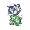 3lf6C 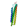 3lf9C 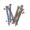 3lg7C 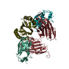 3lh2C 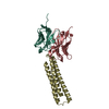 3lhpC 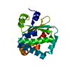 1z6nS C: citing same article ( S: Starting model for refinement |
|---|---|
| Similar structure data |


-Links
 PDBj
PDBj- Assembly
| Deposited unit | 
| |||||||||
|---|---|---|---|---|---|---|---|---|---|---|
| 1 | 
| |||||||||
| Unit cell |
| |||||||||
| Components on special symmetry positions |
|
-Components
| #1: Protein | Mass: 19350.020 Da / Num. of mol.: 1 Source method: isolated from a genetically manipulated source Details: The epitope-scaffold is based on the hypothetical protein PA1234 from Pseudomonas aeruginosa (PDB ID 1Z6N) Source: (gene. exp.) ARTIFICIAL GENE (others) / Plasmid: PET29 / Production host:  |
|---|---|
| #2: Chemical | ChemComp-EDO /   Mass: 62.068 Da / Num. of mol.: 1 / Source method: obtained synthetically / Formula: C2H6O2 Mass: 62.068 Da / Num. of mol.: 1 / Source method: obtained synthetically / Formula: C2H6O2 |
| #3: Water | ChemComp-HOH /  Mass: 18.015 Da / Num. of mol.: 55 / Source method: isolated from a natural source / Formula: H2O Mass: 18.015 Da / Num. of mol.: 55 / Source method: isolated from a natural source / Formula: H2O |
-Experimental details
-Experiment
| Experiment | Method: X-RAY DIFFRACTION / Number of used crystals: 1 |
|---|
- Sample preparation
| Crystal | Density Matthews: 3.85 Å3/Da / Density % sol: 68.03 % |
|---|---|
| Crystal grow | Temperature: 277 K / Method: vapor diffusion, hanging drop / pH: 8.5 Details: Na/K tartrate, Tris, pH 8.5, VAPOR DIFFUSION, HANGING DROP, temperature 277K |
-Data collection
| Diffraction | Mean temperature: 107 K |
|---|---|
| Diffraction source | Source: ROTATING ANODE / Type: RIGAKU MICROMAX-007 / Wavelength: 1.54 Å |
| Detector | Type: RIGAKU RAXIS IV++ / Detector: IMAGE PLATE / Date: Apr 17, 2007 / Details: Rigaku Varimax HR |
| Radiation | Monochromator: Mirrors / Protocol: SINGLE WAVELENGTH / Monochromatic (M) / Laue (L): M / Scattering type: x-ray |
| Radiation wavelength | Wavelength: 1.54 Å / Relative weight: 1 |
| Reflection | Resolution: 2.3→48.3 Å / Num. all: 13770 / Num. obs: 13770 / % possible obs: 93.7 % / Redundancy: 8.4 % / Biso Wilson estimate: 56.3 Å2 / Rmerge(I) obs: 0.086 / Net I/σ(I): 9 |
| Reflection shell | Resolution: 2.3→2.38 Å / Redundancy: 6.83 % / Rmerge(I) obs: 0.373 / Mean I/σ(I) obs: 3.9 / Num. unique all: 1304 / % possible all: 91.5 |
- Processing
| Software |
| |||||||||||||||||||||||||||||||||||||||||||||||||||||||||||||||||||||||||||||||||||||||||||||||||||||||||||||||||||||||||||||
|---|---|---|---|---|---|---|---|---|---|---|---|---|---|---|---|---|---|---|---|---|---|---|---|---|---|---|---|---|---|---|---|---|---|---|---|---|---|---|---|---|---|---|---|---|---|---|---|---|---|---|---|---|---|---|---|---|---|---|---|---|---|---|---|---|---|---|---|---|---|---|---|---|---|---|---|---|---|---|---|---|---|---|---|---|---|---|---|---|---|---|---|---|---|---|---|---|---|---|---|---|---|---|---|---|---|---|---|---|---|---|---|---|---|---|---|---|---|---|---|---|---|---|---|---|---|---|
| Refinement | Method to determine structure: MOLECULAR REPLACEMENT Starting model: Computationally-derived model of the epitope-scaffold, based on 1Z6N Resolution: 2.3→46.78 Å / Cor.coef. Fo:Fc: 0.927 / Cor.coef. Fo:Fc free: 0.937 / SU B: 15.538 / SU ML: 0.189 / Isotropic thermal model: Isotropic / Cross valid method: THROUGHOUT / ESU R: 0.257 / ESU R Free: 0.222 / Stereochemistry target values: MAXIMUM LIKELIHOOD / Details: HYDROGENS HAVE BEEN ADDED IN THE RIDING POSITIONS
| |||||||||||||||||||||||||||||||||||||||||||||||||||||||||||||||||||||||||||||||||||||||||||||||||||||||||||||||||||||||||||||
| Solvent computation | Ion probe radii: 0.8 Å / Shrinkage radii: 0.8 Å / VDW probe radii: 1.4 Å / Solvent model: MASK | |||||||||||||||||||||||||||||||||||||||||||||||||||||||||||||||||||||||||||||||||||||||||||||||||||||||||||||||||||||||||||||
| Displacement parameters | Biso mean: 49.573 Å2
| |||||||||||||||||||||||||||||||||||||||||||||||||||||||||||||||||||||||||||||||||||||||||||||||||||||||||||||||||||||||||||||
| Refinement step | Cycle: LAST / Resolution: 2.3→46.78 Å
| |||||||||||||||||||||||||||||||||||||||||||||||||||||||||||||||||||||||||||||||||||||||||||||||||||||||||||||||||||||||||||||
| Refine LS restraints |
| |||||||||||||||||||||||||||||||||||||||||||||||||||||||||||||||||||||||||||||||||||||||||||||||||||||||||||||||||||||||||||||
| LS refinement shell | Resolution: 2.3→2.36 Å / Total num. of bins used: 20
|
