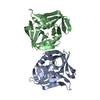
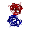
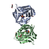
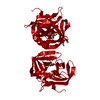
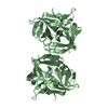
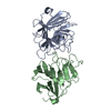
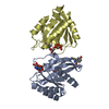
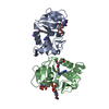
[English] 日本語
Yorodumi
- PDB-3ruo: Complex structure of HevB EV93 main protease 3C with Rupintrivir ... -

+
Open data


ID or keywords:

Loading...

-
Basic information

Entry
Database: PDB / ID: 3ruo
TitleComplex structure of HevB EV93 main protease 3C with Rupintrivir (AG7088)
ComponentsHEVB EV93 3C PROTEASE
KeywordsHYDROLASE/HYDROLASE INHIBITOR / CYSTEINE TRYPSIN-LIKE PROTEASE / 3C CYSTEINE PROTEASE (PICORNAIN 3C) / RUPINTRIVIR (AG7088) / COVALENTLY BOUND INHIBITOR / HYDROLASE-HYDROLASE INHIBITOR complex
Function / homology
Function and homology information


symbiont-mediated suppression of host cytoplasmic pattern recognition receptor signaling pathway via inhibition of RIG-I activity / picornain 2A / symbiont-mediated suppression of host mRNA export from nucleus / symbiont genome entry into host cell via pore formation in plasma membrane / picornain 3C / T=pseudo3 icosahedral viral capsid / host cell cytoplasmic vesicle membrane / cytoplasmic vesicle membrane / endocytosis involved in viral entry into host cell / nucleoside-triphosphate phosphatase ...symbiont-mediated suppression of host cytoplasmic pattern recognition receptor signaling pathway via inhibition of RIG-I activity / picornain 2A / symbiont-mediated suppression of host mRNA export from nucleus / symbiont genome entry into host cell via pore formation in plasma membrane / picornain 3C / T=pseudo3 icosahedral viral capsid / host cell cytoplasmic vesicle membrane / cytoplasmic vesicle membrane / endocytosis involved in viral entry into host cell / nucleoside-triphosphate phosphatase / protein complex oligomerization / monoatomic ion channel activity / symbiont-mediated suppression of host gene expression / DNA replication / RNA helicase activity / induction by virus of host autophagy / RNA-directed RNA polymerase / viral RNA genome replication / cysteine-type endopeptidase activity / RNA-dependent RNA polymerase activity / virus-mediated perturbation of host defense response / DNA-templated transcription / virion attachment to host cell / structural molecule activity / ATP hydrolysis activity / proteolysis / RNA binding / ATP binding / metal ion binding
Similarity search - Function
Picornavirus coat protein VP4 superfamily / Poliovirus 3A protein-like / Poliovirus 3A protein like / Picornavirus 2B protein / Poliovirus core protein 3a, soluble domain / Picornavirus 2B protein / Peptidase C3, picornavirus core protein 2A / Picornavirus core protein 2A / Picornavirus coat protein VP4 / Picornavirus coat protein (VP4) ...Picornavirus coat protein VP4 superfamily / Poliovirus 3A protein-like / Poliovirus 3A protein like / Picornavirus 2B protein / Poliovirus core protein 3a, soluble domain / Picornavirus 2B protein / Peptidase C3, picornavirus core protein 2A / Picornavirus core protein 2A / Picornavirus coat protein VP4 / Picornavirus coat protein (VP4) / Picornavirales 3C/3C-like protease domain / Picornavirales 3C/3C-like protease domain profile. / Peptidase C3A/C3B, picornaviral / 3C cysteine protease (picornain 3C) / Picornavirus capsid / picornavirus capsid protein / Helicase, superfamily 3, single-stranded RNA virus / Superfamily 3 helicase of positive ssRNA viruses domain profile. / Helicase, superfamily 3, single-stranded DNA/RNA virus / RNA helicase / Picornavirus/Calicivirus coat protein / Viral coat protein subunit / Trypsin-like serine proteases / Thrombin, subunit H / RNA-directed RNA polymerase, C-terminal domain / Viral RNA-dependent RNA polymerase / Reverse transcriptase/Diguanylate cyclase domain / RNA-directed RNA polymerase, catalytic domain / RdRp of positive ssRNA viruses catalytic domain profile. / ATPases associated with a variety of cellular activities / AAA+ ATPase domain / Peptidase S1, PA clan, chymotrypsin-like fold / Peptidase S1, PA clan / DNA/RNA polymerase superfamily / Beta Barrel / P-loop containing nucleoside triphosphate hydrolase / Mainly Beta
Similarity search - Domain/homology
Chem-AG7 / Genome polyprotein
Similarity search - Component
Biological speciesHuman enterovirus B
MethodX-RAY DIFFRACTION / SYNCHROTRON / MOLECULAR REPLACEMENT / Resolution: 1.5 Å
AuthorsKaczmarska, Z. / Janowski, R. / Costenaro, L. / Coutard, B. / Norder, H. / Canard, B. / Coll, M.
CitationJournal: J.Virol. / Year: 2011
Title: Structural Basis for Antiviral Inhibition of the Main Protease, 3C, from Human Enterovirus 93.
Authors: Costenaro, L. / Kaczmarska, Z. / Arnan, C. / Janowski, R. / Coutard, B. / Sola, M. / Gorbalenya, A.E. / Norder, H. / Canard, B. / Coll, M.
History
DepositionMay 5, 2011Deposition site: RCSB / Processing site: RCSB
Revision 1.0Sep 7, 2011Provider: repository / Type: Initial release
Revision 1.1Oct 5, 2011Group: Database references
Revision 1.2Sep 13, 2023Group: Data collection / Database references ...Data collection / Database references / Derived calculations / Refinement description
Category: chem_comp_atom / chem_comp_bond ...chem_comp_atom / chem_comp_bond / database_2 / pdbx_initial_refinement_model / pdbx_struct_conn_angle / struct_conn / struct_ref_seq_dif / struct_site
Item: _database_2.pdbx_DOI / _database_2.pdbx_database_accession ..._database_2.pdbx_DOI / _database_2.pdbx_database_accession / _pdbx_struct_conn_angle.ptnr1_auth_asym_id / _pdbx_struct_conn_angle.ptnr1_auth_seq_id / _pdbx_struct_conn_angle.ptnr1_label_asym_id / _pdbx_struct_conn_angle.ptnr2_auth_asym_id / _pdbx_struct_conn_angle.ptnr2_label_asym_id / _pdbx_struct_conn_angle.ptnr3_auth_asym_id / _pdbx_struct_conn_angle.ptnr3_auth_seq_id / _pdbx_struct_conn_angle.ptnr3_label_asym_id / _pdbx_struct_conn_angle.value / _struct_conn.pdbx_dist_value / _struct_conn.pdbx_leaving_atom_flag / _struct_conn.ptnr1_auth_asym_id / _struct_conn.ptnr1_auth_comp_id / _struct_conn.ptnr1_auth_seq_id / _struct_conn.ptnr1_label_asym_id / _struct_conn.ptnr1_label_atom_id / _struct_conn.ptnr1_label_comp_id / _struct_conn.ptnr1_label_seq_id / _struct_conn.ptnr2_auth_asym_id / _struct_conn.ptnr2_auth_comp_id / _struct_conn.ptnr2_auth_seq_id / _struct_conn.ptnr2_label_asym_id / _struct_conn.ptnr2_label_atom_id / _struct_conn.ptnr2_label_comp_id / _struct_conn.ptnr2_label_seq_id / _struct_ref_seq_dif.details / _struct_site.pdbx_auth_asym_id / _struct_site.pdbx_auth_comp_id / _struct_site.pdbx_auth_seq_id

-
Structure visualization

Structure viewerMolecule:
MolmilJmol/JSmol

Downloads & links

-
Assembly

Deposited unit
A: HEVB EV93 3C PROTEASE
B: HEVB EV93 3C PROTEASE
hetero molecules


Theoretical massNumber of molelcules
Total (without water)44,0767
Polymers42,7912
Non-polymers1,2855
Water6,774376
1


  • Idetical with deposited unit
  • defined by author&software
TypeNameSymmetry operationNumber
identity operation1_555x,y,z1
Buried area3840 Å2
ΔGint-39 kcal/mol
Surface area15970 Å2
MethodPISA
Unit cell
Length a, b, c (Å)38.995, 63.911, 66.357
Angle α, β, γ (deg.)90.00, 90.43, 90.00
Int Tables number4
Space group name H-MP1211

-
Components

#1: Protein HEVB EV93 3C PROTEASE


Mass: 21395.512 Da / Num. of mol.: 2
Source method: isolated from a genetically manipulated source
Source: (gene. exp.) Human enterovirus B / Gene: 3C / Plasmid: PDEST14 / Production host: Escherichia coli (E. coli) / Strain (production host): BL21 / References: UniProt: Q5DSM6, picornain 3C
#2: Chemical ChemComp-AG7 / 4-{2-(4-FLUORO-BENZYL)-6-METHYL-5-[(5-METHYL-ISOXAZOLE-3-CARBONYL)-AMINO]-4-OXO-HEPTANOYLAMINO}-5-(2-OXO-PYRROLIDIN-3-YL)-PENTANOIC ACID ETHYL ESTER / RUPINTRIVIR, bound form


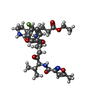
Mass: 600.678 Da / Num. of mol.: 2 / Source method: obtained synthetically / Formula: C31H41FN4O7 / Comment: antivirus, protease inhibitor*YM
#3: Chemical ChemComp-MG / MAGNESIUM ION


Mass: 24.305 Da / Num. of mol.: 2 / Source method: obtained synthetically / Formula: Mg
#4: Chemical ChemComp-CL / CHLORIDE ION


Mass: 35.453 Da / Num. of mol.: 1 / Source method: obtained synthetically / Formula: Cl
#5: Water ChemComp-HOH / water


Mass: 18.015 Da / Num. of mol.: 376 / Source method: isolated from a natural source / Formula: H2O
Sequence detailsAUTHORS STATE THAT THIS MAY BE A NATURAL VARIANT.

-
Experimental details

-
Experiment

ExperimentMethod: X-RAY DIFFRACTION / Number of used crystals: 1

-
Sample preparation

CrystalDensity Matthews: 1.93 Å3/Da / Density % sol: 36.35 %
Crystal growTemperature: 293 K / Method: vapor diffusion, sitting drop / pH: 8.5
Details: 0.25 M MgCl2, 0.1 M Tris, 25% PEG 8k, pH 8.5, VAPOR DIFFUSION, SITTING DROP, temperature 293K

-
Data collection

DiffractionMean temperature: 100 K
Diffraction sourceSource: SYNCHROTRON / Site: ESRF / Beamline: ID23-2 / Wavelength: 0.8726 Å
DetectorType: MARMOSAIC 225 mm CCD / Detector: CCD / Date: Feb 2, 2011 / Details: MIRRORS
RadiationMonochromator: HORIZONTALLY SIDE DIFFRACTING SILICON 111 CRYSTAL
Protocol: SINGLE WAVELENGTH / Monochromatic (M) / Laue (L): M / Scattering type: x-ray
Radiation wavelengthWavelength: 0.8726 Å / Relative weight: 1
ReflectionResolution: 1.5→30 Å / Num. obs: 52513 / % possible obs: 100 % / Observed criterion σ(F): -3 / Observed criterion σ(I): -3
Reflection shellResolution: 1.5→1.55 Å / % possible all: 100

-
Processing

Software
NameVersionClassification
DNAdata collection
MOLREPphasing
REFMAC5.5.0102refinement
DENZOdata reduction
SCALEPACKdata scaling
RefinementMethod to determine structure: MOLECULAR REPLACEMENT
Starting model: PDB entry 3Q3Y
Resolution: 1.5→30 Å / Cor.coef. Fo:Fc: 0.969 / Cor.coef. Fo:Fc free: 0.953 / SU B: 2.851 / SU ML: 0.05 / Cross valid method: THROUGHOUT / σ(F): 0 / σ(I): 0 / ESU R Free: 0.08 / Stereochemistry target values: MAXIMUM LIKELIHOOD
RfactorNum. reflection% reflectionSelection details
Rfree0.19796 2658 5.1 %RANDOM
Rwork0.16581 ---
all0.16745 49523 --
obs0.16745 49523 99.98 %-
Solvent computationIon probe radii: 0.8 Å / Shrinkage radii: 0.8 Å / VDW probe radii: 1.4 Å / Solvent model: BABINET MODEL WITH MASK
Displacement parametersBiso mean: 16.921 Å2
Baniso -1Baniso -2Baniso -3
1--0.24 Å20 Å20.07 Å2
2--0.72 Å20 Å2
3----0.48 Å2
Refinement stepCycle: LAST / Resolution: 1.5→30 Å
ProteinNucleic acidLigandSolventTotal
Num. atoms2842 0 89 376 3307
Refine LS restraints
Refine-IDTypeDev idealDev ideal targetNumber
X-RAY DIFFRACTIONr_bond_refined_d0.0080.0223203
X-RAY DIFFRACTIONr_angle_refined_deg1.4091.9964366
X-RAY DIFFRACTIONr_dihedral_angle_1_deg5.9055418
X-RAY DIFFRACTIONr_dihedral_angle_2_deg31.19623.931145
X-RAY DIFFRACTIONr_dihedral_angle_3_deg11.82815552
X-RAY DIFFRACTIONr_dihedral_angle_4_deg18.1931521
X-RAY DIFFRACTIONr_chiral_restr0.0880.2476
X-RAY DIFFRACTIONr_gen_planes_refined0.0060.0212481
X-RAY DIFFRACTIONr_mcbond_it0.5281.51902
X-RAY DIFFRACTIONr_mcangle_it1.0282.53085
X-RAY DIFFRACTIONr_scbond_it2.32551301
X-RAY DIFFRACTIONr_scangle_it3.733101253
LS refinement shellResolution: 1.5→1.581 Å / Total num. of bins used: 10
RfactorNum. reflection% reflection
Rfree0.256 394 -
Rwork0.223 7138 -
obs--99.97 %
Refinement TLS params.

Method: refined / Refine-ID: X-RAY DIFFRACTION

IDL112)L122)L132)L222)L232)L332)S11 (Å °)S12 (Å °)S13 (Å °)S21 (Å °)S22 (Å °)S23 (Å °)S31 (Å °)S32 (Å °)S33 (Å °)T112)T122)T132)T222)T232)T332)Origin x (Å)Origin y (Å)Origin z (Å)
10.6222-0.417-0.34660.97960.05460.89460.0257-0.08740-0.03460.0166-0.1196-0.02250.0586-0.04230.0154-0.00950.00040.02360.00260.0339-11.150610.8671-0.9255
23.5771-1.02540.06330.95790.81071.2408-0.1251-0.4007-0.01620.12710.08530.00360.1098-0.04520.03980.03590.00460.01030.05430.02310.0416-17.34876.76226.5499
35.9826-2.60112.2892.4323-0.92662.4153-0.0884-0.15380.12170.10480.1041-0.1784-0.21580.1632-0.01570.0389-0.0265-0.00410.0475-0.030.0373-12.723820.44936.1825
41.0593-0.157-0.23620.8559-0.0820.90420.03410.0406-0.0101-0.0698-0.01880.0369-0.0085-0.0763-0.01520.0244-0.0031-0.00440.0108-0.00420.0165-22.037914.5074-7.9087
51.29050.57940.11981.0010.32292.03140.03360.10920.02950.107-0.00010.1233-0.080.001-0.03350.033-0.00260.00470.01680.00440.0489-8.016517.8945-31.7513
61.42940.3570.09730.7569-0.50930.7765-0.03810.311-0.0292-0.06260.04240.00730.03310.0372-0.00430.0159-0.0050.0040.0907-0.03070.0211-3.363413.3948-42.0157
7-0.09110.1285-0.22431.21541.12361.3451-0.0080.12280.2148-0.0867-0.05870.3285-0.2798-0.11430.06660.1722-0.0195-0.02490.1020.07980.2368-7.260527.4642-36.8663
81.68980.1663-0.3130.71220.1391.55770.0356-0.0463-0.02350.0617-0.0135-0.0379-0.00330.1484-0.02210.032-0.006-0.00320.0143-0.00180.01862.448617.677-25.2821
Refinement TLS group
IDRefine-IDRefine TLS-IDAuth asym-IDAuth seq-ID
1X-RAY DIFFRACTION1A-1 - 48
2X-RAY DIFFRACTION2A49 - 78
3X-RAY DIFFRACTION3A79 - 93
4X-RAY DIFFRACTION4A94 - 182
5X-RAY DIFFRACTION5B0 - 40
6X-RAY DIFFRACTION6B41 - 78
7X-RAY DIFFRACTION7B79 - 93
8X-RAY DIFFRACTION8B94 - 180

+
About Yorodumi

-
News

-
Feb 9, 2022. New format data for meta-information of EMDB entries

New format data for meta-information of EMDB entries

  • Version 3 of the EMDB header file is now the official format.
  • The previous official version 1.9 will be removed from the archive.

Related info.:EMDB header

External links:wwPDB to switch to version 3 of the EMDB data model

-
Aug 12, 2020. Covid-19 info

Covid-19 info

URL: https://pdbjlvh1.pdbj.org/emnavi/covid19.php

New page: Covid-19 featured information page in EM Navigator.

Related info.:Covid-19 info / Mar 5, 2020. Novel coronavirus structure data

+
Mar 5, 2020. Novel coronavirus structure data

Novel coronavirus structure data

Related info.:Yorodumi Speices / Aug 12, 2020. Covid-19 info

External links:COVID-19 featured content - PDBj / Molecule of the Month (242):Coronavirus Proteases

+
Jan 31, 2019. EMDB accession codes are about to change! (news from PDBe EMDB page)

EMDB accession codes are about to change! (news from PDBe EMDB page)

  • The allocation of 4 digits for EMDB accession codes will soon come to an end. Whilst these codes will remain in use, new EMDB accession codes will include an additional digit and will expand incrementally as the available range of codes is exhausted. The current 4-digit format prefixed with “EMD-” (i.e. EMD-XXXX) will advance to a 5-digit format (i.e. EMD-XXXXX), and so on. It is currently estimated that the 4-digit codes will be depleted around Spring 2019, at which point the 5-digit format will come into force.
  • The EM Navigator/Yorodumi systems omit the EMD- prefix.

Related info.:Q: What is EMD? / ID/Accession-code notation in Yorodumi/EM Navigator

External links:EMDB Accession Codes are Changing Soon! / Contact to PDBj

+
Jul 12, 2017. Major update of PDB

Major update of PDB

  • wwPDB released updated PDB data conforming to the new PDBx/mmCIF dictionary.
  • This is a major update changing the version number from 4 to 5, and with Remediation, in which all the entries are updated.
  • In this update, many items about electron microscopy experimental information are reorganized (e.g. em_software).
  • Now, EM Navigator and Yorodumi are based on the updated data.

External links:wwPDB Remediation / Enriched Model Files Conforming to OneDep Data Standards Now Available in the PDB FTP Archive

-
Yorodumi

Thousand views of thousand structures

  • Yorodumi is a browser for structure data from EMDB, PDB, SASBDB, etc.
  • This page is also the successor to EM Navigator detail page, and also detail information page/front-end page for Omokage search.
  • The word "yorodu" (or yorozu) is an old Japanese word meaning "ten thousand". "mi" (miru) is to see.

Related info.:EMDB / PDB / SASBDB / Comparison of 3 databanks / Yorodumi Search / Aug 31, 2016. New EM Navigator & Yorodumi / Yorodumi Papers / Jmol/JSmol / Function and homology information / Changes in new EM Navigator and Yorodumi

Read more