 Movie
Movie Controller

[English] 日本語
 Yorodumi
- PDB-3l3h: X-ray crystal structure of the F6A mutant of influenza A acid pol... -
+ Open data

- Basic information
| Entry | Database: PDB / ID: 3l3h | ||||||
|---|---|---|---|---|---|---|---|
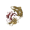
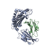
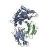
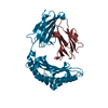
| Title | X-ray crystal structure of the F6A mutant of influenza A acid polymerase epitope PA224 bound to murine H2-Db MHC | ||||||
 Components Components |
| ||||||
 Keywords Keywords | IMMUNE SYSTEM / Animals Antigens / Viral Epitopes / T-Lymphocyte Histocompatibility Antigens / Mice Antigen / T-Cell / T-Lymphocytes / Glycoprotein / Immune response / Membrane / MHC I / Transmembrane / Immunoglobulin domain / Secreted | ||||||
| Function / homology |  Function and homology information Function and homology informationEndosomal/Vacuolar pathway / DAP12 interactions / Antigen Presentation: Folding, assembly and peptide loading of class I MHC / ER-Phagosome pathway / DAP12 signaling / Immunoregulatory interactions between a Lymphoid and a non-Lymphoid cell / cap snatching / regulation of membrane depolarization / symbiont-mediated suppression of host mRNA transcription via inhibition of RNA polymerase II activity / antigen processing and presentation of peptide antigen via MHC class I ...Endosomal/Vacuolar pathway / DAP12 interactions / Antigen Presentation: Folding, assembly and peptide loading of class I MHC / ER-Phagosome pathway / DAP12 signaling / Immunoregulatory interactions between a Lymphoid and a non-Lymphoid cell / cap snatching / regulation of membrane depolarization / symbiont-mediated suppression of host mRNA transcription via inhibition of RNA polymerase II activity / antigen processing and presentation of peptide antigen via MHC class I / cellular defense response / Neutrophil degranulation / lumenal side of endoplasmic reticulum membrane / regulation of iron ion transport / cellular response to iron(III) ion / antigen processing and presentation of exogenous protein antigen via MHC class Ib, TAP-dependent / negative regulation of iron ion transport / negative regulation of forebrain neuron differentiation / regulation of erythrocyte differentiation / iron ion transport / peptide antigen assembly with MHC class I protein complex / response to molecule of bacterial origin / HFE-transferrin receptor complex / MHC class I peptide loading complex / transferrin transport / negative regulation of receptor-mediated endocytosis / cellular response to iron ion / positive regulation of T cell cytokine production / antigen processing and presentation of endogenous peptide antigen via MHC class I / MHC class I protein complex / peptide antigen assembly with MHC class II protein complex / negative regulation of neurogenesis / multicellular organismal-level iron ion homeostasis / cellular response to nicotine / MHC class II protein complex / positive regulation of receptor-mediated endocytosis / positive regulation of T cell mediated cytotoxicity / negative regulation of epithelial cell proliferation / antigen processing and presentation of exogenous peptide antigen via MHC class II / positive regulation of immune response / peptide antigen binding / phagocytic vesicle membrane / positive regulation of T cell activation / sensory perception of smell / positive regulation of cellular senescence / MHC class II protein complex binding / T cell differentiation in thymus / late endosome membrane / negative regulation of neuron projection development / antimicrobial humoral immune response mediated by antimicrobial peptide / antibacterial humoral response / cellular response to lipopolysaccharide / protein refolding / endonuclease activity / amyloid fibril formation / defense response to Gram-negative bacterium / protein homotetramerization / Hydrolases; Acting on ester bonds / intracellular iron ion homeostasis / host cell cytoplasm / learning or memory / defense response to Gram-positive bacterium / immune response / symbiont-mediated suppression of host gene expression / external side of plasma membrane / viral translational frameshifting / innate immune response / lysosomal membrane / viral RNA genome replication / hydrolase activity / host cell nucleus / structural molecule activity / Golgi apparatus / DNA-templated transcription / protein homodimerization activity / : / RNA binding / metal ion binding / identical protein binding / plasma membrane / cytosol Similarity search - Function | ||||||
| Biological species |  | ||||||
| Method |  X-RAY DIFFRACTION / MOLECULAR REPLACEMENT / Resolution: 2.7 Å X-RAY DIFFRACTION / MOLECULAR REPLACEMENT / Resolution: 2.7 Å | ||||||
 Authors Authors | Welland, A. / Clements, C.S. / Dunstone, M.A. / Rossjohn, J. | ||||||
 Citation Citation | Journal: Proc.Natl.Acad.Sci.USA / Year: 2010 Title: Constraints within major histocompatibility complex class I restricted peptides: presentation and consequences for T-cell recognition Authors: Theodossis, A. / Guillonneau, C. / Welland, A. / Ely, L.K. / Clements, C.S. / Williamson, N.A. / Webb, A.I. / Wilce, J.A. / Mulder, R.J. / Dunstone, M.A. / Doherty, P.C. / McCluskey, J. / ...Authors: Theodossis, A. / Guillonneau, C. / Welland, A. / Ely, L.K. / Clements, C.S. / Williamson, N.A. / Webb, A.I. / Wilce, J.A. / Mulder, R.J. / Dunstone, M.A. / Doherty, P.C. / McCluskey, J. / Purcell, A.W. / Turner, S.J. / Rossjohn, J. | ||||||
| History |
|
- Structure visualization
| Structure viewer | Molecule: MolmilJmol/JSmol |
|---|
- Downloads & links
-Download
| PDBx/mmCIF format | 3l3h.cif.gz | 91.9 KB | Display | PDBx/mmCIF format |
|---|---|---|---|---|
| PDB format | pdb3l3h.ent.gz | 69.4 KB | Display | PDB format |
| PDBx/mmJSON format | 3l3h.json.gz | Tree view | PDBx/mmJSON format | |
| Others |  Other downloads Other downloads |
-Validation report
| Arichive directory | https://data.pdbj.org/pub/pdb/validation_reports/l3/3l3hftp://data.pdbj.org/pub/pdb/validation_reports/l3/3l3h | HTTPS FTP |
|---|
-Related structure data
| Related structure data |  3l3dC  3l3gC  3l3iC  3l3jC  3l3kC  3cc5S C: citing same article ( S: Starting model for refinement |
|---|---|
| Similar structure data |


-Links
 PDBj
PDBj


- Assembly
| Deposited unit | 
| ||||||||
|---|---|---|---|---|---|---|---|---|---|
| 1 |
| ||||||||
| Unit cell |
|
-Components
| #1: Protein | Mass: 32030.648 Da / Num. of mol.: 1 Source method: isolated from a genetically manipulated source Source: (gene. exp.)  |
|---|---|
| #2: Protein | Mass: 11704.359 Da / Num. of mol.: 1 Source method: isolated from a genetically manipulated source Source: (gene. exp.) |
| #3: Protein/peptide | Mass: 1110.199 Da / Num. of mol.: 1 / Mutation: F6A / Source method: obtained synthetically / Details: Fmoc-peptide synthesis / References: UniProt: Q17TI0 |
| #4: Water | ChemComp-HOH /  Mass: 18.015 Da / Num. of mol.: 122 / Source method: isolated from a natural source / Formula: H2O Mass: 18.015 Da / Num. of mol.: 122 / Source method: isolated from a natural source / Formula: H2O |
| Has protein modification | Y |
-Experimental details
-Experiment
| Experiment | Method: X-RAY DIFFRACTION / Number of used crystals: 1 |
|---|
- Sample preparation
| Crystal | Density Matthews: 2.41 Å3/Da / Density % sol: 48.98 % |
|---|---|
| Crystal grow | Temperature: 293 K / Method: vapor diffusion, hanging drop / pH: 5.6 Details: 0.1M citrate, 0.2M ammonium acetate, 25% PEG4000, pH 5.6, VAPOR DIFFUSION, HANGING DROP, temperature 293K |
-Data collection
| Diffraction | Mean temperature: 100 K |
|---|---|
| Diffraction source | Source: ROTATING ANODE / Type: RIGAKU / Wavelength: 1.51478 Å |
| Detector | Type: RIGAKU RAXIS IV++ / Detector: IMAGE PLATE / Date: Apr 2, 2004 |
| Radiation | Protocol: SINGLE WAVELENGTH / Monochromatic (M) / Laue (L): M / Scattering type: x-ray |
| Radiation wavelength | Wavelength: 1.51478 Å / Relative weight: 1 |
| Reflection | Resolution: 2.7→28 Å / Num. all: 12799 / Num. obs: 12799 / % possible obs: 98.2 % / Observed criterion σ(F): 1 / Observed criterion σ(I): 1 / Redundancy: 4.5 % / Rmerge(I) obs: 0.091 / Net I/σ(I): 18.4 |
| Reflection shell | Resolution: 2.7→2.8 Å / Redundancy: 4.6 % / Rmerge(I) obs: 0.317 / Mean I/σ(I) obs: 5.1 / % possible all: 98 |
- Processing
| Software |
| ||||||||||||||||||||||||||||||||||||||||||||||||||||||||||||||||||||||||||||||||||||||||||||||||||||
|---|---|---|---|---|---|---|---|---|---|---|---|---|---|---|---|---|---|---|---|---|---|---|---|---|---|---|---|---|---|---|---|---|---|---|---|---|---|---|---|---|---|---|---|---|---|---|---|---|---|---|---|---|---|---|---|---|---|---|---|---|---|---|---|---|---|---|---|---|---|---|---|---|---|---|---|---|---|---|---|---|---|---|---|---|---|---|---|---|---|---|---|---|---|---|---|---|---|---|---|---|---|
| Refinement | Method to determine structure: MOLECULAR REPLACEMENT Starting model: 3CC5 Resolution: 2.7→28 Å / Cor.coef. Fo:Fc: 0.904 / Cor.coef. Fo:Fc free: 0.854 / SU B: 27.379 / SU ML: 0.295 / TLS residual ADP flag: LIKELY RESIDUAL / Cross valid method: THROUGHOUT / ESU R Free: 0.426 / Stereochemistry target values: MAXIMUM LIKELIHOOD / Details: HYDROGENS HAVE BEEN ADDED IN THE RIDING POSITIONS
| ||||||||||||||||||||||||||||||||||||||||||||||||||||||||||||||||||||||||||||||||||||||||||||||||||||
| Solvent computation | Ion probe radii: 0.8 Å / Shrinkage radii: 0.8 Å / VDW probe radii: 1.2 Å / Solvent model: MASK | ||||||||||||||||||||||||||||||||||||||||||||||||||||||||||||||||||||||||||||||||||||||||||||||||||||
| Displacement parameters | Biso mean: 22.889 Å2
| ||||||||||||||||||||||||||||||||||||||||||||||||||||||||||||||||||||||||||||||||||||||||||||||||||||
| Refinement step | Cycle: LAST / Resolution: 2.7→28 Å
| ||||||||||||||||||||||||||||||||||||||||||||||||||||||||||||||||||||||||||||||||||||||||||||||||||||
| Refine LS restraints |
| ||||||||||||||||||||||||||||||||||||||||||||||||||||||||||||||||||||||||||||||||||||||||||||||||||||
| LS refinement shell | Resolution: 2.701→2.771 Å / Total num. of bins used: 20
| ||||||||||||||||||||||||||||||||||||||||||||||||||||||||||||||||||||||||||||||||||||||||||||||||||||
| Refinement TLS params. | Method: refined / Refine-ID: X-RAY DIFFRACTION
| ||||||||||||||||||||||||||||||||||||||||||||||||||||||||||||||||||||||||||||||||||||||||||||||||||||
| Refinement TLS group |
|
