 Movie
Movie Controller

[English] 日本語
 Yorodumi
- PDB-3l3k: Crystal structure of HLA-B*4402 in complex with the R5A/F7A doubl... -
+ Open data

- Basic information
| Entry | Database: PDB / ID: 3l3k | ||||||
|---|---|---|---|---|---|---|---|
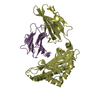
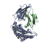
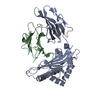
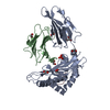
| Title | Crystal structure of HLA-B*4402 in complex with the R5A/F7A double mutant of a self-peptide derived from DPA*0201 | ||||||
 Components Components |
| ||||||
 Keywords Keywords | IMMUNE SYSTEM / Immunoglobulin domain / Immune response / Major Histocompatibility Complex Class I / MHC-I peptide complex / altered peptide ligand | ||||||
| Function / homology |  Function and homology information Function and homology informationantigen processing and presentation of peptide or polysaccharide antigen via MHC class II / regulation of interleukin-12 production / regulation of dendritic cell differentiation / regulation of T cell anergy / regulation of interleukin-6 production / protection from natural killer cell mediated cytotoxicity / TAP binding / detection of bacterium / antigen processing and presentation of endogenous peptide antigen via MHC class Ib / antigen processing and presentation of endogenous peptide antigen via MHC class I via ER pathway, TAP-independent ...antigen processing and presentation of peptide or polysaccharide antigen via MHC class II / regulation of interleukin-12 production / regulation of dendritic cell differentiation / regulation of T cell anergy / regulation of interleukin-6 production / protection from natural killer cell mediated cytotoxicity / TAP binding / detection of bacterium / antigen processing and presentation of endogenous peptide antigen via MHC class Ib / antigen processing and presentation of endogenous peptide antigen via MHC class I via ER pathway, TAP-independent / secretory granule membrane / early endosome lumen / Nef mediated downregulation of MHC class I complex cell surface expression / DAP12 interactions / Endosomal/Vacuolar pathway / T cell mediated cytotoxicity / Antigen Presentation: Folding, assembly and peptide loading of class I MHC / lumenal side of endoplasmic reticulum membrane / regulation of iron ion transport / cellular response to iron(III) ion / negative regulation of iron ion transport / negative regulation of forebrain neuron differentiation / antigen processing and presentation of exogenous protein antigen via MHC class Ib, TAP-dependent / peptide antigen assembly with MHC class I protein complex / ER to Golgi transport vesicle membrane / regulation of erythrocyte differentiation / response to molecule of bacterial origin / HFE-transferrin receptor complex / defense response / MHC class I peptide loading complex / transferrin transport / cellular response to iron ion / negative regulation of receptor-mediated endocytosis / positive regulation of T cell cytokine production / antigen processing and presentation of endogenous peptide antigen via MHC class I / MHC class I protein complex / peptide antigen assembly with MHC class II protein complex / negative regulation of neurogenesis / MHC class II protein complex / cellular response to nicotine / positive regulation of receptor-mediated endocytosis / multicellular organismal-level iron ion homeostasis / positive regulation of T cell mediated cytotoxicity / specific granule lumen / antigen processing and presentation of exogenous peptide antigen via MHC class II / positive regulation of immune response / peptide antigen binding / phagocytic vesicle membrane / recycling endosome membrane / positive regulation of T cell activation / negative regulation of epithelial cell proliferation / Interferon gamma signaling / Immunoregulatory interactions between a Lymphoid and a non-Lymphoid cell / Interferon alpha/beta signaling / sensory perception of smell / Modulation by Mtb of host immune system / positive regulation of cellular senescence / tertiary granule lumen / MHC class II protein complex binding / T cell differentiation in thymus / DAP12 signaling / late endosome membrane / negative regulation of neuron projection development / protein refolding / protein-folding chaperone binding / ER-Phagosome pathway / early endosome membrane / amyloid fibril formation / protein homotetramerization / intracellular iron ion homeostasis / adaptive immune response / learning or memory / endosome membrane / immune response / endoplasmic reticulum lumen / Amyloid fiber formation / signaling receptor binding / Golgi membrane / external side of plasma membrane / innate immune response / lysosomal membrane / focal adhesion / Neutrophil degranulation / SARS-CoV-2 activates/modulates innate and adaptive immune responses / structural molecule activity / cell surface / Golgi apparatus / endoplasmic reticulum / protein homodimerization activity / : / extracellular exosome / extracellular region / membrane / identical protein binding / plasma membrane / cytosol Similarity search - Function | ||||||
| Biological species |  Homo sapiens (human) Homo sapiens (human) | ||||||
| Method |  X-RAY DIFFRACTION / SYNCHROTRON / MOLECULAR REPLACEMENT / Resolution: 2.6 Å X-RAY DIFFRACTION / SYNCHROTRON / MOLECULAR REPLACEMENT / Resolution: 2.6 Å | ||||||
 Authors Authors | Theodossis, A. / Ely, L.K. / Rossjohn, J. | ||||||
 Citation Citation | Journal: Proc.Natl.Acad.Sci.USA / Year: 2010 Title: Constraints within major histocompatibility complex class I restricted peptides: presentation and consequences for T-cell recognition Authors: Theodossis, A. / Guillonneau, C. / Welland, A. / Ely, L.K. / Clements, C.S. / Williamson, N.A. / Webb, A.I. / Wilce, J.A. / Mulder, R.J. / Dunstone, M.A. / Doherty, P.C. / McCluskey, J. / ...Authors: Theodossis, A. / Guillonneau, C. / Welland, A. / Ely, L.K. / Clements, C.S. / Williamson, N.A. / Webb, A.I. / Wilce, J.A. / Mulder, R.J. / Dunstone, M.A. / Doherty, P.C. / McCluskey, J. / Purcell, A.W. / Turner, S.J. / Rossjohn, J. | ||||||
| History |
|
- Structure visualization
| Structure viewer | Molecule: MolmilJmol/JSmol |
|---|
- Downloads & links
-Download
| PDBx/mmCIF format | 3l3k.cif.gz | 92.2 KB | Display | PDBx/mmCIF format |
|---|---|---|---|---|
| PDB format | pdb3l3k.ent.gz | 69.8 KB | Display | PDB format |
| PDBx/mmJSON format | 3l3k.json.gz | Tree view | PDBx/mmJSON format | |
| Others |  Other downloads Other downloads |
-Validation report
| Arichive directory | https://data.pdbj.org/pub/pdb/validation_reports/l3/3l3kftp://data.pdbj.org/pub/pdb/validation_reports/l3/3l3k | HTTPS FTP |
|---|
-Related structure data
| Related structure data |  3l3dC  3l3gC  3l3hC  3l3iC  3l3jC  1m6oS S: Starting model for refinement C: citing same article ( |
|---|---|
| Similar structure data |


-Links
 PDBj
PDBj


- Assembly
| Deposited unit | 
| ||||||||
|---|---|---|---|---|---|---|---|---|---|
| 1 |
| ||||||||
| Unit cell |
|
-Components
| #1: Protein | Mass: 31980.258 Da / Num. of mol.: 1 / Fragment: extracellular domain Source method: isolated from a genetically manipulated source Source: (gene. exp.) Homo sapiens (human) / Gene: HLA-B, HLAB / Production host:  |
|---|---|
| #2: Protein | Mass: 11748.160 Da / Num. of mol.: 1 Source method: isolated from a genetically manipulated source Source: (gene. exp.) Homo sapiens (human) / Gene: B2M, CDABP0092, HDCMA22P / Production host: |
| #3: Protein/peptide | Mass: 927.953 Da / Num. of mol.: 1 / Mutation: R5A,F7A / Source method: obtained synthetically / Details: automated Fmoc-peptide synthesis / References: UniProt: Q95HB9 |
| #4: Chemical | ChemComp-GOL /   Mass: 92.094 Da / Num. of mol.: 1 / Source method: obtained synthetically / Formula: C3H8O3 Mass: 92.094 Da / Num. of mol.: 1 / Source method: obtained synthetically / Formula: C3H8O3 |
| #5: Water | ChemComp-HOH /  Mass: 18.015 Da / Num. of mol.: 142 / Source method: isolated from a natural source / Formula: H2O Mass: 18.015 Da / Num. of mol.: 142 / Source method: isolated from a natural source / Formula: H2O |
| Has protein modification | Y |
-Experimental details
-Experiment
| Experiment | Method: X-RAY DIFFRACTION / Number of used crystals: 1 |
|---|
- Sample preparation
| Crystal | Density Matthews: 2.54 Å3/Da / Density % sol: 51.55 % |
|---|---|
| Crystal grow | Temperature: 293 K / Method: vapor diffusion, hanging drop / pH: 5.5 Details: 0.1M tri-sodium citrate dihydrate, 12-30% PEG 4000, 0.2M ammonium acetate, pH 5.5, VAPOR DIFFUSION, HANGING DROP, temperature 293K |
-Data collection
| Diffraction | Mean temperature: 100 K |
|---|---|
| Diffraction source | Source: SYNCHROTRON / Site: APS  / Beamline: 14-BM-C / Beamline: 14-BM-C |
| Detector | Type: ADSC QUANTUM 4 / Detector: CCD / Date: Nov 20, 2002 |
| Radiation | Protocol: SINGLE WAVELENGTH / Monochromatic (M) / Laue (L): M / Scattering type: x-ray |
| Radiation wavelength | Relative weight: 1 |
| Reflection | Resolution: 2.5→65.6 Å / Num. all: 15875 / Num. obs: 15875 / % possible obs: 93.6 % / Observed criterion σ(F): 1 / Observed criterion σ(I): 1 / Redundancy: 2.7 % / Rmerge(I) obs: 0.132 / Net I/σ(I): 10.6 |
| Reflection shell | Resolution: 2.5→2.6 Å / Rmerge(I) obs: 0.245 / Mean I/σ(I) obs: 3 / % possible all: 71.3 |
- Processing
| Software |
| |||||||||||||||||||||||||||||||||||||||||||||||||||||||||||||||||||||||||||||||||||||||||||||||||||||||||||||||||||||||||||||
|---|---|---|---|---|---|---|---|---|---|---|---|---|---|---|---|---|---|---|---|---|---|---|---|---|---|---|---|---|---|---|---|---|---|---|---|---|---|---|---|---|---|---|---|---|---|---|---|---|---|---|---|---|---|---|---|---|---|---|---|---|---|---|---|---|---|---|---|---|---|---|---|---|---|---|---|---|---|---|---|---|---|---|---|---|---|---|---|---|---|---|---|---|---|---|---|---|---|---|---|---|---|---|---|---|---|---|---|---|---|---|---|---|---|---|---|---|---|---|---|---|---|---|---|---|---|---|
| Refinement | Method to determine structure: MOLECULAR REPLACEMENT Starting model: PDB ENTRY 1M6O Resolution: 2.6→25 Å / Cor.coef. Fo:Fc: 0.918 / Cor.coef. Fo:Fc free: 0.878 / SU B: 26.516 / SU ML: 0.256 / TLS residual ADP flag: LIKELY RESIDUAL Isotropic thermal model: isotropic atomic temperature factors with TLS Cross valid method: THROUGHOUT / ESU R Free: 0.38 / Stereochemistry target values: MAXIMUM LIKELIHOOD
| |||||||||||||||||||||||||||||||||||||||||||||||||||||||||||||||||||||||||||||||||||||||||||||||||||||||||||||||||||||||||||||
| Solvent computation | Ion probe radii: 0.8 Å / Shrinkage radii: 0.8 Å / VDW probe radii: 1.4 Å / Solvent model: MASK | |||||||||||||||||||||||||||||||||||||||||||||||||||||||||||||||||||||||||||||||||||||||||||||||||||||||||||||||||||||||||||||
| Displacement parameters | Biso mean: 10.712 Å2
| |||||||||||||||||||||||||||||||||||||||||||||||||||||||||||||||||||||||||||||||||||||||||||||||||||||||||||||||||||||||||||||
| Refinement step | Cycle: LAST / Resolution: 2.6→25 Å
| |||||||||||||||||||||||||||||||||||||||||||||||||||||||||||||||||||||||||||||||||||||||||||||||||||||||||||||||||||||||||||||
| Refine LS restraints |
| |||||||||||||||||||||||||||||||||||||||||||||||||||||||||||||||||||||||||||||||||||||||||||||||||||||||||||||||||||||||||||||
| LS refinement shell | Resolution: 2.6→2.667 Å / Total num. of bins used: 20
| |||||||||||||||||||||||||||||||||||||||||||||||||||||||||||||||||||||||||||||||||||||||||||||||||||||||||||||||||||||||||||||
| Refinement TLS params. | Method: refined / Refine-ID: X-RAY DIFFRACTION
| |||||||||||||||||||||||||||||||||||||||||||||||||||||||||||||||||||||||||||||||||||||||||||||||||||||||||||||||||||||||||||||
| Refinement TLS group |
|
