 Movie
Movie Controller

[English] 日本語
 Yorodumi
- PDB-3khj: C. parvum inosine monophosphate dehydrogenase bound by inhibitor C64 -
+ Open data

- Basic information
| Entry | Database: PDB / ID: 3khj | ||||||
|---|---|---|---|---|---|---|---|
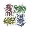
| Title | C. parvum inosine monophosphate dehydrogenase bound by inhibitor C64 | ||||||
 Components Components | Inosine-5-monophosphate dehydrogenase | ||||||
 Keywords Keywords | OXIDOREDUCTASE / enzyme-inhibitor complex | ||||||
| Function / homology |  Function and homology information Function and homology informationIMP dehydrogenase / IMP dehydrogenase activity / GMP biosynthetic process / GTP biosynthetic process Similarity search - Function | ||||||
| Biological species |   Cryptosporidium parvum (eukaryote) Cryptosporidium parvum (eukaryote) | ||||||
| Method |  X-RAY DIFFRACTION / SYNCHROTRON / MOLECULAR REPLACEMENT / Resolution: 2.8 Å X-RAY DIFFRACTION / SYNCHROTRON / MOLECULAR REPLACEMENT / Resolution: 2.8 Å | ||||||
 Authors Authors | MacPherson, I.S. / Hedstrom, L.K. | ||||||
 Citation Citation | Journal: J.Am.Chem.Soc. / Year: 2010 Title: The structural basis of Cryptosporidium -specific IMP dehydrogenase inhibitor selectivity. Authors: Macpherson, I.S. / Kirubakaran, S. / Gorla, S.K. / Riera, T.V. / D'Aquino, J.A. / Zhang, M. / Cuny, G.D. / Hedstrom, L. | ||||||
| History |
|
- Structure visualization
| Structure viewer | Molecule: MolmilJmol/JSmol |
|---|
- Downloads & links
-Download
| PDBx/mmCIF format | 3khj.cif.gz | 451.8 KB | Display | PDBx/mmCIF format |
|---|---|---|---|---|
| PDB format | pdb3khj.ent.gz | 366.6 KB | Display | PDB format |
| PDBx/mmJSON format | 3khj.json.gz | Tree view | PDBx/mmJSON format | |
| Others |  Other downloads Other downloads |
-Validation report
| Arichive directory | https://data.pdbj.org/pub/pdb/validation_reports/kh/3khjftp://data.pdbj.org/pub/pdb/validation_reports/kh/3khj | HTTPS FTP |
|---|
-Related structure data


-Links
 PDBj
PDBj- Assembly
| Deposited unit | 
| ||||||||
|---|---|---|---|---|---|---|---|---|---|
| 1 | 
| ||||||||
| 2 | 
| ||||||||
| Unit cell |
| ||||||||
| Details | Tetramer |
-Components
| #1: Protein | Mass: 38516.441 Da / Num. of mol.: 8 Source method: isolated from a genetically manipulated source Source: (gene. exp.) Cryptosporidium parvum (eukaryote) / Strain: Iowa-II / Gene: cgd6_20, GuaB / Plasmid: pET28a / Production host:  #2: Chemical | ChemComp-IMP /   Mass: 348.206 Da / Num. of mol.: 8 / Source method: obtained synthetically / Formula: C10H13N4O8P Mass: 348.206 Da / Num. of mol.: 8 / Source method: obtained synthetically / Formula: C10H13N4O8P#3: Chemical |   Mass: 413.291 Da / Num. of mol.: 3 / Source method: obtained synthetically / Formula: C18H13BrN4OS Mass: 413.291 Da / Num. of mol.: 3 / Source method: obtained synthetically / Formula: C18H13BrN4OS#4: Chemical | ChemComp-ACT / |   Mass: 59.044 Da / Num. of mol.: 1 / Source method: obtained synthetically / Formula: C2H3O2 Mass: 59.044 Da / Num. of mol.: 1 / Source method: obtained synthetically / Formula: C2H3O2#5: Water | ChemComp-HOH / |  Mass: 18.015 Da / Num. of mol.: 208 / Source method: isolated from a natural source / Formula: H2O Mass: 18.015 Da / Num. of mol.: 208 / Source method: isolated from a natural source / Formula: H2O |
|---|
-Experimental details
-Experiment
| Experiment | Method: X-RAY DIFFRACTION / Number of used crystals: 1 |
|---|
- Sample preparation
| Crystal | Density Matthews: 2.2 Å3/Da / Density % sol: 44.1 % |
|---|---|
| Crystal grow | Temperature: 295 K / Method: microbatch under oil / pH: 4.6 Details: 30% MPD, 100 mM sodium acetate pH 4.6, 20 mM calcium chloride, microbatch under oil, temperature 295K |
-Data collection
| Diffraction | Mean temperature: 100 K |
|---|---|
| Diffraction source | Source: SYNCHROTRON / Site: ALS  / Beamline: 8.2.2 / Wavelength: 0.9194 Å / Beamline: 8.2.2 / Wavelength: 0.9194 Å |
| Detector | Type: ADSC QUANTUM 315 / Detector: CCD / Date: Oct 30, 2008 |
| Radiation | Monochromator: Double crystal Si(111) / Protocol: SINGLE WAVELENGTH / Monochromatic (M) / Laue (L): M / Scattering type: x-ray |
| Radiation wavelength | Wavelength: 0.9194 Å / Relative weight: 1 |
| Reflection | Resolution: 2.8→100 Å / Num. all: 65589 / Num. obs: 65589 / % possible obs: 100 % / Observed criterion σ(F): 1 / Observed criterion σ(I): 1 |
| Reflection shell | Resolution: 2.8→2.9 Å / Rmerge(I) obs: 0.383 / Mean I/σ(I) obs: 5.2 / % possible all: 100 |
- Processing
| Software |
| ||||||||||||||||||||||||||||||||||||||||||||||||||||||||||||||||||||||||||||||||||||||||||||||||||||||||||||||||||||||||||||||||||||||||||||||||||||||||||||||||||||||||||
|---|---|---|---|---|---|---|---|---|---|---|---|---|---|---|---|---|---|---|---|---|---|---|---|---|---|---|---|---|---|---|---|---|---|---|---|---|---|---|---|---|---|---|---|---|---|---|---|---|---|---|---|---|---|---|---|---|---|---|---|---|---|---|---|---|---|---|---|---|---|---|---|---|---|---|---|---|---|---|---|---|---|---|---|---|---|---|---|---|---|---|---|---|---|---|---|---|---|---|---|---|---|---|---|---|---|---|---|---|---|---|---|---|---|---|---|---|---|---|---|---|---|---|---|---|---|---|---|---|---|---|---|---|---|---|---|---|---|---|---|---|---|---|---|---|---|---|---|---|---|---|---|---|---|---|---|---|---|---|---|---|---|---|---|---|---|---|---|---|---|---|---|
| Refinement | Method to determine structure: MOLECULAR REPLACEMENT / Resolution: 2.8→42.135 Å / Cor.coef. Fo:Fc: 0.919 / Cor.coef. Fo:Fc free: 0.885 / SU B: 16.103 / SU ML: 0.319 / Cross valid method: THROUGHOUT / ESU R Free: 0.415 / Stereochemistry target values: MAXIMUM LIKELIHOOD / Details: HYDROGENS HAVE BEEN ADDED IN THE RIDING POSITIONS
| ||||||||||||||||||||||||||||||||||||||||||||||||||||||||||||||||||||||||||||||||||||||||||||||||||||||||||||||||||||||||||||||||||||||||||||||||||||||||||||||||||||||||||
| Solvent computation | Ion probe radii: 0.8 Å / Shrinkage radii: 0.8 Å / VDW probe radii: 1.2 Å / Solvent model: MASK | ||||||||||||||||||||||||||||||||||||||||||||||||||||||||||||||||||||||||||||||||||||||||||||||||||||||||||||||||||||||||||||||||||||||||||||||||||||||||||||||||||||||||||
| Displacement parameters | Biso mean: 53.214 Å2
| ||||||||||||||||||||||||||||||||||||||||||||||||||||||||||||||||||||||||||||||||||||||||||||||||||||||||||||||||||||||||||||||||||||||||||||||||||||||||||||||||||||||||||
| Refinement step | Cycle: LAST / Resolution: 2.8→42.135 Å
| ||||||||||||||||||||||||||||||||||||||||||||||||||||||||||||||||||||||||||||||||||||||||||||||||||||||||||||||||||||||||||||||||||||||||||||||||||||||||||||||||||||||||||
| Refine LS restraints |
| ||||||||||||||||||||||||||||||||||||||||||||||||||||||||||||||||||||||||||||||||||||||||||||||||||||||||||||||||||||||||||||||||||||||||||||||||||||||||||||||||||||||||||
| LS refinement shell | Resolution: 2.796→2.869 Å / Total num. of bins used: 20
|
