 Movie
Movie Controller

[English] 日本語
 Yorodumi
- PDB-3ffs: The Crystal Structure of Cryptosporidium parvum Inosine-5'-Monoph... -
+ Open data

- Basic information
| Entry | Database: PDB / ID: 3ffs | ||||||
|---|---|---|---|---|---|---|---|
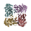
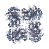
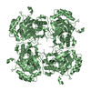
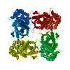
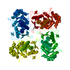
| Title | The Crystal Structure of Cryptosporidium parvum Inosine-5'-Monophosphate Dehydrogenase | ||||||
 Components Components | Inosine-5-monophosphate dehydrogenase | ||||||
 Keywords Keywords | OXIDOREDUCTASE / beta-alpha barrel / TIM fold | ||||||
| Function / homology |  Function and homology information Function and homology informationIMP dehydrogenase / IMP dehydrogenase activity / GMP biosynthetic process / GTP biosynthetic process / metal ion binding / cytoplasm Similarity search - Function | ||||||
| Biological species |   Cryptosporidium parvum (eukaryote) Cryptosporidium parvum (eukaryote) | ||||||
| Method |  X-RAY DIFFRACTION / SYNCHROTRON / MOLECULAR REPLACEMENT / molecular replacement / Resolution: 3.19 Å X-RAY DIFFRACTION / SYNCHROTRON / MOLECULAR REPLACEMENT / molecular replacement / Resolution: 3.19 Å | ||||||
 Authors Authors | Riera, T.V. / D'Aquino, J.A. / Lu, J. / Petsko, G.A. / Hedstrom, L. | ||||||
 Citation Citation | Journal: J.Am.Chem.Soc. / Year: 2010 Title: The structural basis of Cryptosporidium -specific IMP dehydrogenase inhibitor selectivity Authors: Macpherson, I.S. / Kirubakaran, S. / Gorla, S.K. / Riera, T.V. / D'Aquino, J.A. / Zhang, M. / Cuny, G.D. / Hedstrom, L. | ||||||
| History |
|
- Structure visualization
| Structure viewer | Molecule: MolmilJmol/JSmol |
|---|
- Downloads & links
-Download
| PDBx/mmCIF format | 3ffs.cif.gz | 221.1 KB | Display | PDBx/mmCIF format |
|---|---|---|---|---|
| PDB format | pdb3ffs.ent.gz | 175.8 KB | Display | PDB format |
| PDBx/mmJSON format | 3ffs.json.gz | Tree view | PDBx/mmJSON format | |
| Others |  Other downloads Other downloads |
-Validation report
| Arichive directory | https://data.pdbj.org/pub/pdb/validation_reports/ff/3ffsftp://data.pdbj.org/pub/pdb/validation_reports/ff/3ffs | HTTPS FTP |
|---|
-Related structure data
| Related structure data |  3khjC  1eepS S: Starting model for refinement C: citing same article ( |
|---|---|
| Similar structure data |

-Links
 PDBj
PDBj- Assembly
| Deposited unit | 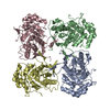
| ||||||||||||||||||||||||||||||||||||||||||||||||||||||||||||||||||||||||||||||||||||||||||||||||||||||||||||||||||||||||||||
|---|---|---|---|---|---|---|---|---|---|---|---|---|---|---|---|---|---|---|---|---|---|---|---|---|---|---|---|---|---|---|---|---|---|---|---|---|---|---|---|---|---|---|---|---|---|---|---|---|---|---|---|---|---|---|---|---|---|---|---|---|---|---|---|---|---|---|---|---|---|---|---|---|---|---|---|---|---|---|---|---|---|---|---|---|---|---|---|---|---|---|---|---|---|---|---|---|---|---|---|---|---|---|---|---|---|---|---|---|---|---|---|---|---|---|---|---|---|---|---|---|---|---|---|---|---|
| 1 |
| ||||||||||||||||||||||||||||||||||||||||||||||||||||||||||||||||||||||||||||||||||||||||||||||||||||||||||||||||||||||||||||
| Unit cell |
| ||||||||||||||||||||||||||||||||||||||||||||||||||||||||||||||||||||||||||||||||||||||||||||||||||||||||||||||||||||||||||||
| Noncrystallographic symmetry (NCS) | NCS domain:
NCS domain segments: Component-ID: 1 / Beg auth comp-ID: THR / Beg label comp-ID: THR / End auth comp-ID: THR / End label comp-ID: THR / Refine code: 6 / Auth seq-ID: 3 - 378 / Label seq-ID: 3 - 378
NCS ensembles :
|
-Components
| #1: Protein | Mass: 43134.262 Da / Num. of mol.: 4 Source method: isolated from a genetically manipulated source Source: (gene. exp.) Cryptosporidium parvum (eukaryote) / Gene: IMPDH, 56k.02 / Plasmid: pTacTac / Production host:  |
|---|
-Experimental details
-Experiment
| Experiment | Method: X-RAY DIFFRACTION / Number of used crystals: 1 |
|---|
- Sample preparation
| Crystal | Density Matthews: 2.6 Å3/Da / Density % sol: 52.66 % |
|---|---|
| Crystal grow | Temperature: 298 K / Method: vapor diffusion, hanging drop / pH: 8.5 Details: Protein solution: 4 mg/mL IMPDH, 50 mM Tris, pH 7.5, 150 mM KCl, 5 % glycerol and 2 mM dithiothreitol. Well solution: 34 % PEG 4000, 25 mM sodium acetate and 100 mM Tris-Cl, pH 8.5. Combined ...Details: Protein solution: 4 mg/mL IMPDH, 50 mM Tris, pH 7.5, 150 mM KCl, 5 % glycerol and 2 mM dithiothreitol. Well solution: 34 % PEG 4000, 25 mM sodium acetate and 100 mM Tris-Cl, pH 8.5. Combined in a 1:1 ratio. VAPOR DIFFUSION, HANGING DROP, temperature 298K |
-Data collection
| Diffraction | Mean temperature: 100 K |
|---|---|
| Diffraction source | Source: SYNCHROTRON / Site: APS  / Beamline: 8-BM / Wavelength: 0.97946 Å / Beamline: 8-BM / Wavelength: 0.97946 Å |
| Detector | Type: ADSC QUANTUM 315 / Detector: CCD / Date: Oct 27, 2006 |
| Radiation | Monochromator: Double Crystal Monochromator / Protocol: SINGLE WAVELENGTH / Monochromatic (M) / Laue (L): M / Scattering type: x-ray |
| Radiation wavelength | Wavelength: 0.97946 Å / Relative weight: 1 |
| Reflection | Resolution: 3.19→50 Å / Num. obs: 30575 / % possible obs: 100 % / Redundancy: 6.8 % / Rmerge(I) obs: 0.179 / Χ2: 1.171 |
| Reflection shell | Resolution: 3.19→3.31 Å / Redundancy: 6.9 % / Rmerge(I) obs: 0.632 / Num. unique all: 2987 / Χ2: 1.072 / % possible all: 100 |
-Phasing
| Phasing | Method: molecular replacement |
|---|
- Processing
| Software |
| |||||||||||||||||||||||||||||||||||||||||||||||||||||||||||||||||||||||||||||||||||||||||||||||||||||||||||||||||||||||||||||||||||||||||||||||||||||||||||||||||||||||||||
|---|---|---|---|---|---|---|---|---|---|---|---|---|---|---|---|---|---|---|---|---|---|---|---|---|---|---|---|---|---|---|---|---|---|---|---|---|---|---|---|---|---|---|---|---|---|---|---|---|---|---|---|---|---|---|---|---|---|---|---|---|---|---|---|---|---|---|---|---|---|---|---|---|---|---|---|---|---|---|---|---|---|---|---|---|---|---|---|---|---|---|---|---|---|---|---|---|---|---|---|---|---|---|---|---|---|---|---|---|---|---|---|---|---|---|---|---|---|---|---|---|---|---|---|---|---|---|---|---|---|---|---|---|---|---|---|---|---|---|---|---|---|---|---|---|---|---|---|---|---|---|---|---|---|---|---|---|---|---|---|---|---|---|---|---|---|---|---|---|---|---|---|---|
| Refinement | Method to determine structure: MOLECULAR REPLACEMENT Starting model: PDB entry 1EEP Resolution: 3.19→48.34 Å / Cor.coef. Fo:Fc: 0.866 / Cor.coef. Fo:Fc free: 0.82 / WRfactor Rfree: 0.313 / WRfactor Rwork: 0.259 / Occupancy max: 1 / Occupancy min: 1 / FOM work R set: 0.953 / SU B: 26.148 / SU ML: 0.45 / SU Rfree: 0.58 / Cross valid method: THROUGHOUT / ESU R Free: 0.58 Stereochemistry target values: MAXIMUM LIKELIHOOD WITH PHASES
| |||||||||||||||||||||||||||||||||||||||||||||||||||||||||||||||||||||||||||||||||||||||||||||||||||||||||||||||||||||||||||||||||||||||||||||||||||||||||||||||||||||||||||
| Solvent computation | Ion probe radii: 0.8 Å / Shrinkage radii: 0.8 Å / VDW probe radii: 1.4 Å / Solvent model: MASK | |||||||||||||||||||||||||||||||||||||||||||||||||||||||||||||||||||||||||||||||||||||||||||||||||||||||||||||||||||||||||||||||||||||||||||||||||||||||||||||||||||||||||||
| Displacement parameters | Biso max: 84.19 Å2 / Biso mean: 53.887 Å2 / Biso min: 20 Å2
| |||||||||||||||||||||||||||||||||||||||||||||||||||||||||||||||||||||||||||||||||||||||||||||||||||||||||||||||||||||||||||||||||||||||||||||||||||||||||||||||||||||||||||
| Refinement step | Cycle: LAST / Resolution: 3.19→48.34 Å
| |||||||||||||||||||||||||||||||||||||||||||||||||||||||||||||||||||||||||||||||||||||||||||||||||||||||||||||||||||||||||||||||||||||||||||||||||||||||||||||||||||||||||||
| Refine LS restraints |
| |||||||||||||||||||||||||||||||||||||||||||||||||||||||||||||||||||||||||||||||||||||||||||||||||||||||||||||||||||||||||||||||||||||||||||||||||||||||||||||||||||||||||||
| Refine LS restraints NCS | Number: 1614 / Refine-ID: X-RAY DIFFRACTION
|