 Movie
Movie Controller

[English] 日本語
 Yorodumi
- PDB-1mei: Inosine Monophosphate Dehydrogenase (IMPDH) From Tritrichomonas F... -
+ Open data

- Basic information
| Entry | Database: PDB / ID: 1mei | |||||||||
|---|---|---|---|---|---|---|---|---|---|---|
| Title | Inosine Monophosphate Dehydrogenase (IMPDH) From Tritrichomonas Foetus with XMP and mycophenolic acid bound | |||||||||
 Components Components | INOSINE-5'-MONOPHOSPHATE DEHYDROGENASE | |||||||||
 Keywords Keywords | OXIDOREDUCTASE / alpha beta barrel | |||||||||
| Function / homology |  Function and homology information Function and homology informationIMP dehydrogenase / IMP dehydrogenase activity / GMP biosynthetic process / GTP biosynthetic process / nucleotide binding / protein-containing complex / metal ion binding / identical protein binding / cytoplasm Similarity search - Function | |||||||||
| Biological species |  Tritrichomonas foetus (eukaryote) Tritrichomonas foetus (eukaryote) | |||||||||
| Method |  X-RAY DIFFRACTION / SYNCHROTRON / FOURIER SYNTHESIS / Resolution: 2.2 Å X-RAY DIFFRACTION / SYNCHROTRON / FOURIER SYNTHESIS / Resolution: 2.2 Å | |||||||||
 Authors Authors | Prosise, G.L. / Luecke, H. | |||||||||
 Citation Citation | Journal: J.Mol.Biol. / Year: 2003 Title: Crystal Structures of Tritrichomonas foetus Inosine Monophosphate Dehydrogenase in Complex with Substrate, Cofactor and Analogs: A Structural Basis for the Random-in Ordered-out Kinetic Mechanism Authors: Prosise, G.L. / Luecke, H. | |||||||||
| History |
|
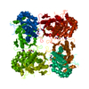
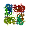
- Structure visualization
| Structure viewer | Molecule: MolmilJmol/JSmol |
|---|
- Downloads & links
-Download
| PDBx/mmCIF format | 1mei.cif.gz | 89.9 KB | Display | PDBx/mmCIF format |
|---|---|---|---|---|
| PDB format | pdb1mei.ent.gz | 65.6 KB | Display | PDB format |
| PDBx/mmJSON format | 1mei.json.gz | Tree view | PDBx/mmJSON format | |
| Others |  Other downloads Other downloads |
-Validation report
| Arichive directory | https://data.pdbj.org/pub/pdb/validation_reports/me/1meiftp://data.pdbj.org/pub/pdb/validation_reports/me/1mei | HTTPS FTP |
|---|
-Related structure data
| Related structure data |  1me9C  1mehC  1mewC 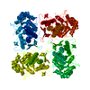 1ak5S S: Starting model for refinement C: citing same article ( |
|---|---|
| Similar structure data |


-Links
 PDBj
PDBj
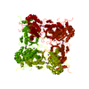
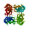
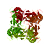
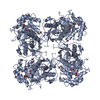
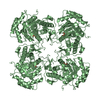
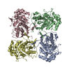
- Assembly
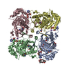
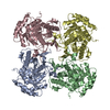
| Deposited unit | 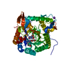
| ||||||||
|---|---|---|---|---|---|---|---|---|---|
| 1 | 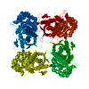
| ||||||||
| Unit cell |
| ||||||||
| Components on special symmetry positions |
|
-Components
| #1: Protein | Mass: 55540.012 Da / Num. of mol.: 1 Source method: isolated from a genetically manipulated source Source: (gene. exp.) Tritrichomonas foetus (eukaryote) / Gene: IMPDH / Plasmid: pBACE / Production host:  |
|---|---|
| #2: Chemical | ChemComp-K /   Mass: 39.098 Da / Num. of mol.: 1 / Source method: obtained synthetically / Formula: K Mass: 39.098 Da / Num. of mol.: 1 / Source method: obtained synthetically / Formula: K |
| #3: Chemical | ChemComp-XMP / 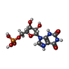  Mass: 365.213 Da / Num. of mol.: 1 / Source method: obtained synthetically / Formula: C10H14N4O9P Mass: 365.213 Da / Num. of mol.: 1 / Source method: obtained synthetically / Formula: C10H14N4O9P |
| #4: Chemical | ChemComp-MOA / 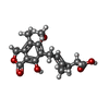  Mass: 320.337 Da / Num. of mol.: 1 / Source method: obtained synthetically / Formula: C17H20O6 Mass: 320.337 Da / Num. of mol.: 1 / Source method: obtained synthetically / Formula: C17H20O6 |
| #5: Water | ChemComp-HOH /  Mass: 18.015 Da / Num. of mol.: 180 / Source method: isolated from a natural source / Formula: H2O Mass: 18.015 Da / Num. of mol.: 180 / Source method: isolated from a natural source / Formula: H2O |
| Has protein modification | Y |
-Experimental details
-Experiment
| Experiment | Method: X-RAY DIFFRACTION / Number of used crystals: 1 |
|---|
- Sample preparation
| Crystal | Density Matthews: 2.8 Å3/Da / Density % sol: 56.02 % | |||||||||||||||||||||||||||||||||||||||||||||||||||||||||||||||||||||||||||||
|---|---|---|---|---|---|---|---|---|---|---|---|---|---|---|---|---|---|---|---|---|---|---|---|---|---|---|---|---|---|---|---|---|---|---|---|---|---|---|---|---|---|---|---|---|---|---|---|---|---|---|---|---|---|---|---|---|---|---|---|---|---|---|---|---|---|---|---|---|---|---|---|---|---|---|---|---|---|---|
| Crystal grow | Temperature: 293 K / Method: vapor diffusion, sitting drop / pH: 7.5 Details: sodium malonate, Tris, 2-mercaptoethanol, EDTA, glycerol, pH 7.5, VAPOR DIFFUSION, SITTING DROP, temperature 293K | |||||||||||||||||||||||||||||||||||||||||||||||||||||||||||||||||||||||||||||
| Crystal grow | *PLUS Temperature: 20 ℃ / pH: 8 / Method: vapor diffusion, sitting drop | |||||||||||||||||||||||||||||||||||||||||||||||||||||||||||||||||||||||||||||
| Components of the solutions | *PLUS
|
-Data collection
| Diffraction | Mean temperature: 100 K |
|---|---|
| Diffraction source | Source: SYNCHROTRON / Site: SSRL  / Beamline: BL9-1 / Wavelength: 0.97 Å / Beamline: BL9-1 / Wavelength: 0.97 Å |
| Detector | Type: MARRESEARCH / Detector: IMAGE PLATE / Date: Apr 11, 2001 |
| Radiation | Protocol: SINGLE WAVELENGTH / Monochromatic (M) / Laue (L): M / Scattering type: x-ray |
| Radiation wavelength | Wavelength: 0.97 Å / Relative weight: 1 |
| Reflection | Resolution: 2.2→99 Å / Num. all: 32987 / Num. obs: 32815 / % possible obs: 99.5 % / Redundancy: 4.7 % / Biso Wilson estimate: 27.4 Å2 / Rmerge(I) obs: 0.066 / Net I/σ(I): 21.4 |
| Reflection shell | Resolution: 2.2→2.24 Å / Rmerge(I) obs: 0.5 / Mean I/σ(I) obs: 2.9 / Num. unique all: 1622 / % possible all: 98.7 |
| Reflection | *PLUS Highest resolution: 2.2 Å / Lowest resolution: 50 Å / Num. obs: 32987 / % possible obs: 99.5 % / Num. measured all: 407857 / Rmerge(I) obs: 0.066 |
| Reflection shell | *PLUS Highest resolution: 2.2 Å / Lowest resolution: 2.34 Å / % possible obs: 98.7 % / Rmerge(I) obs: 0.538 / Mean I/σ(I) obs: 2.88 |
- Processing
| Software |
| ||||||||||||||||||||||||||||||||||||||||||||||||||||||||||||||||||||||||||||||||
|---|---|---|---|---|---|---|---|---|---|---|---|---|---|---|---|---|---|---|---|---|---|---|---|---|---|---|---|---|---|---|---|---|---|---|---|---|---|---|---|---|---|---|---|---|---|---|---|---|---|---|---|---|---|---|---|---|---|---|---|---|---|---|---|---|---|---|---|---|---|---|---|---|---|---|---|---|---|---|---|---|---|
| Refinement | Method to determine structure: FOURIER SYNTHESIS Starting model: pdb entry 1AK5 Resolution: 2.2→33.06 Å / Rfactor Rfree error: 0.006 / Data cutoff high absF: 3510188.21 / Data cutoff low absF: 0 / Isotropic thermal model: RESTRAINED / Cross valid method: THROUGHOUT / σ(F): 0 / σ(I): 0 / Stereochemistry target values: Engh & Huber
| ||||||||||||||||||||||||||||||||||||||||||||||||||||||||||||||||||||||||||||||||
| Solvent computation | Solvent model: FLAT MODEL / Bsol: 35.4457 Å2 / ksol: 0.344434 e/Å3 | ||||||||||||||||||||||||||||||||||||||||||||||||||||||||||||||||||||||||||||||||
| Displacement parameters | Biso mean: 37.3 Å2
| ||||||||||||||||||||||||||||||||||||||||||||||||||||||||||||||||||||||||||||||||
| Refine analyze |
| ||||||||||||||||||||||||||||||||||||||||||||||||||||||||||||||||||||||||||||||||
| Refinement step | Cycle: LAST / Resolution: 2.2→33.06 Å
| ||||||||||||||||||||||||||||||||||||||||||||||||||||||||||||||||||||||||||||||||
| Refine LS restraints |
| ||||||||||||||||||||||||||||||||||||||||||||||||||||||||||||||||||||||||||||||||
| LS refinement shell | Resolution: 2.2→2.34 Å / Rfactor Rfree error: 0.018 / Total num. of bins used: 6
| ||||||||||||||||||||||||||||||||||||||||||||||||||||||||||||||||||||||||||||||||
| Xplor file |
| ||||||||||||||||||||||||||||||||||||||||||||||||||||||||||||||||||||||||||||||||
| Refinement | *PLUS Highest resolution: 2.2 Å / Lowest resolution: 50 Å / Rfactor Rfree: 0.256 / Rfactor Rwork: 0.226 | ||||||||||||||||||||||||||||||||||||||||||||||||||||||||||||||||||||||||||||||||
| Solvent computation | *PLUS | ||||||||||||||||||||||||||||||||||||||||||||||||||||||||||||||||||||||||||||||||
| Displacement parameters | *PLUS | ||||||||||||||||||||||||||||||||||||||||||||||||||||||||||||||||||||||||||||||||
| Refine LS restraints | *PLUS
| ||||||||||||||||||||||||||||||||||||||||||||||||||||||||||||||||||||||||||||||||
| LS refinement shell | *PLUS Highest resolution: 2.2 Å |
