 Movie
Movie Controller

[English] 日本語
 Yorodumi
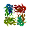
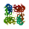
- PDB-1lrt: CRYSTAL STRUCTURE OF TERNARY COMPLEX OF TRITRICHOMONAS FOETUS INO... -
+ Open data

- Basic information
| Entry | Database: PDB / ID: 1lrt | ||||||
|---|---|---|---|---|---|---|---|
| Title | CRYSTAL STRUCTURE OF TERNARY COMPLEX OF TRITRICHOMONAS FOETUS INOSINE-5'-MONOPHOSPHATE DEHYDROGENASE: STRUCTURAL CHARACTERIZATION OF NAD+ SITE IN MICROBIAL ENZYME | ||||||
 Components Components | INOSINE-5'-MONOPHOSPHATE DEHYDROGENASE | ||||||
 Keywords Keywords | OXIDOREDUCTASE / ternary complex / alpha-beta barrel / flexible loop / flap | ||||||
| Function / homology |  Function and homology information Function and homology informationIMP dehydrogenase / IMP dehydrogenase activity / GMP biosynthetic process / GTP biosynthetic process / nucleotide binding / protein-containing complex / metal ion binding / identical protein binding / cytoplasm Similarity search - Function | ||||||
| Biological species |  Tritrichomonas foetus (eukaryote) Tritrichomonas foetus (eukaryote) | ||||||
| Method |  X-RAY DIFFRACTION / SYNCHROTRON / MOLECULAR REPLACEMENT / Resolution: 2.2 Å X-RAY DIFFRACTION / SYNCHROTRON / MOLECULAR REPLACEMENT / Resolution: 2.2 Å | ||||||
 Authors Authors | Gan, L. / Petsko, G.A. / Hedstrom, L. | ||||||
 Citation Citation | Journal: Biochemistry / Year: 2003 Title: Crystal structure of a ternary complex of Tritrichomonas foetus inosine 5'-monophosphate dehydrogenase: NAD+ orients the active site loop for catalysis Authors: Gan, L. / Petsko, G.A. / Hedstrom, L. | ||||||
| History |
| ||||||
| Remark 999 | SEQUENCE AUTHOR STATES THE WILD TYPE T. FOETUS IMPDH CONSISTS OF 503 RESIDUES, HOWEVER THEIR ...SEQUENCE AUTHOR STATES THE WILD TYPE T. FOETUS IMPDH CONSISTS OF 503 RESIDUES, HOWEVER THEIR PROTEIN WAS A MUTANT OFIMPDH. RESIDUES 101-226 IN THE SUBDOMAIN WERE DELETED BECAUSE IT IS NOT REQUIRED FOR IMPDH ACTIVITY. ACCORDING TO THE AUTHORS THE SUBDOMAIN WAS DELETED IN THIS CONSTRUCT TO FACILITATE CRYSTALLIZATION AND THERE IS NO EXOGENOUS LINKER BETWEEN THE RESIDUES 100 AND 227. AUTHOR ALSO STATES ALTHOUGH RESIDUE 279 IS GLU IN SWISS DATABANK, BOTH SEQUENCING RESULT AND ELECTRON DENSITY OF THE STRUCTURE SHOW RESIDUE 279 AS ASP IN THEIR PROTEIN. THE AUTHOR SUGGEST A CONSERVED MUTATION OCCURS AFTER THE CDNA OF THE ENZYME GENE IS TRANSFORMED INTO E. COLI. |
- Structure visualization
| Structure viewer | Molecule: MolmilJmol/JSmol |
|---|
- Downloads & links
-Download
| PDBx/mmCIF format | 1lrt.cif.gz | 289 KB | Display | PDBx/mmCIF format |
|---|---|---|---|---|
| PDB format | pdb1lrt.ent.gz | 231.5 KB | Display | PDB format |
| PDBx/mmJSON format | 1lrt.json.gz | Tree view | PDBx/mmJSON format | |
| Others |  Other downloads Other downloads |
-Validation report
| Arichive directory | https://data.pdbj.org/pub/pdb/validation_reports/lr/1lrtftp://data.pdbj.org/pub/pdb/validation_reports/lr/1lrt | HTTPS FTP |
|---|
-Related structure data
| Related structure data | 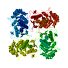 1ak5S S: Starting model for refinement |
|---|---|
| Similar structure data |

-Links
 PDBj
PDBj
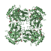
- Assembly
| Deposited unit | 
| ||||||||
|---|---|---|---|---|---|---|---|---|---|
| 1 |
| ||||||||
| Unit cell |
|
-Components
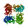
-Protein / Sugars , 2 types, 6 molecules ABCD
| #1: Protein | Mass: 41198.891 Da / Num. of mol.: 4 Source method: isolated from a genetically manipulated source Source: (gene. exp.) Tritrichomonas foetus (eukaryote) / Gene: IMPDH / Plasmid: pKK233-3 / Production host:  #2: Sugar |  Type: D-saccharide / Mass: 292.369 Da / Num. of mol.: 2 Type: D-saccharide / Mass: 292.369 Da / Num. of mol.: 2Source method: isolated from a genetically manipulated source Formula: C14H28O6 / Comment: detergent*YM |
|---|
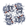
-Non-polymers , 5 types, 567 molecules 


| #3: Chemical | ChemComp-K /  Mass: 39.098 Da / Num. of mol.: 4 / Source method: obtained synthetically / Formula: K Mass: 39.098 Da / Num. of mol.: 4 / Source method: obtained synthetically / Formula: K#4: Chemical | ChemComp-IMP /  Mass: 348.206 Da / Num. of mol.: 4 / Source method: obtained synthetically / Formula: C10H13N4O8P Mass: 348.206 Da / Num. of mol.: 4 / Source method: obtained synthetically / Formula: C10H13N4O8P#5: Chemical | ChemComp-TAD /  Mass: 667.480 Da / Num. of mol.: 4 / Source method: obtained synthetically / Formula: C20H27N7O13P2S Mass: 667.480 Da / Num. of mol.: 4 / Source method: obtained synthetically / Formula: C20H27N7O13P2S#6: Chemical | ChemComp-TRS /  Mass: 122.143 Da / Num. of mol.: 12 / Source method: obtained synthetically / Formula: C4H12NO3 / Comment: pH buffer*YM Mass: 122.143 Da / Num. of mol.: 12 / Source method: obtained synthetically / Formula: C4H12NO3 / Comment: pH buffer*YM#7: Water | ChemComp-HOH / | Mass: 18.015 Da / Num. of mol.: 543 / Source method: isolated from a natural source / Formula: H2O |
|---|
-Details
| Has protein modification | Y |
|---|
-Experimental details
-Experiment
| Experiment | Method: X-RAY DIFFRACTION / Number of used crystals: 1 |
|---|
- Sample preparation
| Crystal | Density Matthews: 2.72 Å3/Da / Density % sol: 54 % | |||||||||||||||||||||||||||||||||||||||||||||||||||||||||||||||||||||||||||||||||||||||||||
|---|---|---|---|---|---|---|---|---|---|---|---|---|---|---|---|---|---|---|---|---|---|---|---|---|---|---|---|---|---|---|---|---|---|---|---|---|---|---|---|---|---|---|---|---|---|---|---|---|---|---|---|---|---|---|---|---|---|---|---|---|---|---|---|---|---|---|---|---|---|---|---|---|---|---|---|---|---|---|---|---|---|---|---|---|---|---|---|---|---|---|---|---|
| Crystal grow | Temperature: 298 K / Method: vapor diffusion, sitting drop / pH: 6.25 Details: PEG 10,000, MES, glycerol, potassium chloride, beta-octylglucoside, pH 6.25, VAPOR DIFFUSION, SITTING DROP, temperature 298.0K | |||||||||||||||||||||||||||||||||||||||||||||||||||||||||||||||||||||||||||||||||||||||||||
| Crystal grow | *PLUS pH: 7.5 / Method: vapor diffusion, sitting drop | |||||||||||||||||||||||||||||||||||||||||||||||||||||||||||||||||||||||||||||||||||||||||||
| Components of the solutions | *PLUS
|
-Data collection
| Diffraction | Mean temperature: 100 K |
|---|---|
| Diffraction source | Source: SYNCHROTRON / Site: APS  / Beamline: 14-BM-C / Wavelength: 1 Å / Beamline: 14-BM-C / Wavelength: 1 Å |
| Detector | Type: ADSC QUANTUM 4 / Detector: CCD / Date: Mar 5, 2001 |
| Radiation | Protocol: SINGLE WAVELENGTH / Monochromatic (M) / Laue (L): M / Scattering type: x-ray |
| Radiation wavelength | Wavelength: 1 Å / Relative weight: 1 |
| Reflection | Resolution: 2.2→30 Å / Num. all: 87015 / Num. obs: 82096 / % possible obs: 97.7 % / Observed criterion σ(F): 0 / Observed criterion σ(I): 1 / Redundancy: 4.1 % / Biso Wilson estimate: 19.3 Å2 / Rmerge(I) obs: 0.089 / Rsym value: 0.089 / Net I/σ(I): 12.2 |
| Reflection shell | Resolution: 2.2→2.28 Å / Redundancy: 2.7 % / Rmerge(I) obs: 0.398 / Mean I/σ(I) obs: 3.4 / Num. unique all: 8087 / Rsym value: 0.398 / % possible all: 91.3 |
| Reflection | *PLUS Num. obs: 87015 / % possible obs: 96.8 % / Num. measured all: 356029 |
| Reflection shell | *PLUS % possible obs: 91.3 % |
- Processing
| Software |
| ||||||||||||||||||||||||||||||||||||||||||||||||||||||
|---|---|---|---|---|---|---|---|---|---|---|---|---|---|---|---|---|---|---|---|---|---|---|---|---|---|---|---|---|---|---|---|---|---|---|---|---|---|---|---|---|---|---|---|---|---|---|---|---|---|---|---|---|---|---|---|
| Refinement | Method to determine structure: MOLECULAR REPLACEMENT Starting model: PDB ENTRY 1AK5 Resolution: 2.2→29.86 Å / Rfactor Rfree error: 0.003 / Data cutoff high absF: 392538.27 / Data cutoff high rms absF: 392538.27 / Data cutoff low absF: 0 / Isotropic thermal model: RESTRAINED / Cross valid method: THROUGHOUT / σ(F): 0 / Stereochemistry target values: Engh & Huber
| ||||||||||||||||||||||||||||||||||||||||||||||||||||||
| Solvent computation | Solvent model: FLAT MODEL / Bsol: 69.6917 Å2 / ksol: 0.421356 e/Å3 | ||||||||||||||||||||||||||||||||||||||||||||||||||||||
| Displacement parameters | Biso mean: 33.7 Å2
| ||||||||||||||||||||||||||||||||||||||||||||||||||||||
| Refine analyze |
| ||||||||||||||||||||||||||||||||||||||||||||||||||||||
| Refinement step | Cycle: LAST / Resolution: 2.2→29.86 Å
| ||||||||||||||||||||||||||||||||||||||||||||||||||||||
| Refine LS restraints |
| ||||||||||||||||||||||||||||||||||||||||||||||||||||||
| LS refinement shell | Refine-ID: X-RAY DIFFRACTION / Total num. of bins used: 6
| ||||||||||||||||||||||||||||||||||||||||||||||||||||||
| Xplor file |
| ||||||||||||||||||||||||||||||||||||||||||||||||||||||
| Refinement | *PLUS Highest resolution: 2.2 Å / Lowest resolution: 8 Å / Num. reflection obs: 71997 / Num. reflection Rfree: 8033 / Rfactor Rwork: 0.218 | ||||||||||||||||||||||||||||||||||||||||||||||||||||||
| Solvent computation | *PLUS | ||||||||||||||||||||||||||||||||||||||||||||||||||||||
| Displacement parameters | *PLUS | ||||||||||||||||||||||||||||||||||||||||||||||||||||||
| Refine LS restraints | *PLUS
|
