 Movie
Movie Controller

[English] 日本語
 Yorodumi
- PDB-3kbm: Room Temperature X-ray structure of D-Xylose Isomerase complexed ... -
+ Open data

- Basic information
| Entry | Database: PDB / ID: 3kbm | ||||||
|---|---|---|---|---|---|---|---|
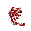
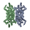
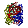
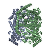
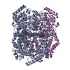
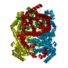
| Title | Room Temperature X-ray structure of D-Xylose Isomerase complexed with 2Cd(2+) co-factors and d12-D-alpha-glucose in the cyclic form | ||||||
 Components Components | Xylose isomerase | ||||||
 Keywords Keywords | ISOMERASE / xylose isomerase / cyclic glucose / Carbohydrate metabolism / Metal-binding / Pentose shunt / Xylose metabolism | ||||||
| Function / homology |  Function and homology information Function and homology informationxylose isomerase / xylose isomerase activity / D-xylose metabolic process / magnesium ion binding / identical protein binding / cytoplasm Similarity search - Function | ||||||
| Biological species |  Streptomyces rubiginosus (bacteria) Streptomyces rubiginosus (bacteria) | ||||||
| Method |  X-RAY DIFFRACTION / MOLECULAR REPLACEMENT / Resolution: 2 Å X-RAY DIFFRACTION / MOLECULAR REPLACEMENT / Resolution: 2 Å | ||||||
 Authors Authors | Kovalevsky, A.Y. / Hanson, L. / Langan, P. | ||||||
 Citation Citation | Journal: Structure / Year: 2010 Title: Metal ion roles and the movement of hydrogen during reaction catalyzed by D-xylose isomerase: a joint x-ray and neutron diffraction study. Authors: Kovalevsky, A.Y. / Hanson, L. / Fisher, S.Z. / Mustyakimov, M. / Mason, S.A. / Forsyth, V.T. / Blakeley, M.P. / Keen, D.A. / Wagner, T. / Carrell, H.L. / Katz, A.K. / Glusker, J.P. / Langan, P. | ||||||
| History |
|
- Structure visualization
| Structure viewer | Molecule: MolmilJmol/JSmol |
|---|
- Downloads & links
-Download
| PDBx/mmCIF format | 3kbm.cif.gz | 96.3 KB | Display | PDBx/mmCIF format |
|---|---|---|---|---|
| PDB format | pdb3kbm.ent.gz | 71.2 KB | Display | PDB format |
| PDBx/mmJSON format | 3kbm.json.gz | Tree view | PDBx/mmJSON format | |
| Others |  Other downloads Other downloads |
-Validation report
| Arichive directory | https://data.pdbj.org/pub/pdb/validation_reports/kb/3kbmftp://data.pdbj.org/pub/pdb/validation_reports/kb/3kbm | HTTPS FTP |
|---|
-Related structure data
| Related structure data | 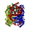 3kbnC 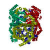 3kbsC 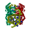 3kbvC  3kbwC 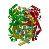 3kclC 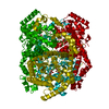 3kcoC 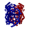 1xibS C: citing same article ( S: Starting model for refinement |
|---|---|
| Similar structure data |


-Links
 PDBj
PDBj


- Assembly
| Deposited unit | 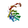
| |||||||||
|---|---|---|---|---|---|---|---|---|---|---|
| 1 | 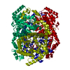
| |||||||||
| Unit cell |
| |||||||||
| Components on special symmetry positions |
|
-Components
| #1: Protein | Mass: 43283.297 Da / Num. of mol.: 1 Source method: isolated from a genetically manipulated source Details: THE PROTEIN WAS PURCHASED FROM HAMPTON RESEARCH / Source: (gene. exp.) Streptomyces rubiginosus (bacteria) / Gene: xylA / References: UniProt: P24300, xylose isomerase | ||||||
|---|---|---|---|---|---|---|---|
| #2: Chemical |   Mass: 112.411 Da / Num. of mol.: 2 / Source method: obtained synthetically / Formula: Cd Mass: 112.411 Da / Num. of mol.: 2 / Source method: obtained synthetically / Formula: Cd#3: Sugar | ChemComp-GLC / |   Type: D-saccharide, alpha linking / Mass: 180.156 Da / Num. of mol.: 1 Type: D-saccharide, alpha linking / Mass: 180.156 Da / Num. of mol.: 1Source method: isolated from a genetically manipulated source Formula: C6H12O6 #4: Water | ChemComp-HOH / |  Mass: 18.015 Da / Num. of mol.: 210 / Source method: isolated from a natural source / Formula: H2O Mass: 18.015 Da / Num. of mol.: 210 / Source method: isolated from a natural source / Formula: H2OSource details | THE PROTEIN WAS PURCHASED FROM HAMPTON RESEARCH | |
-Experimental details
-Experiment
| Experiment | Method: X-RAY DIFFRACTION / Number of used crystals: 1 |
|---|
- Sample preparation
| Crystal | Density Matthews: 2.79 Å3/Da / Density % sol: 55.84 % |
|---|---|
| Crystal grow | Temperature: 290 K / pH: 7.7 Details: 40mg/ml protein, 5mM CdCl2; 500mM d12-D-glucose, 30% (v/v) ammonium sulfate (sat.), batch, pH 7.7, temperature 290K |
-Data collection
| Diffraction | Mean temperature: 293 K |
|---|---|
| Diffraction source | Source: ROTATING ANODE / Type: RIGAKU FR-E DW / Wavelength: 1.54 Å |
| Detector | Type: RIGAKU RAXIS IV++ / Detector: IMAGE PLATE / Date: Jan 15, 2009 / Details: Varimax mirrors |
| Radiation | Monochromator: mirrors / Protocol: SINGLE WAVELENGTH / Monochromatic (M) / Laue (L): M / Scattering type: x-ray |
| Radiation wavelength | Wavelength: 1.54 Å / Relative weight: 1 |
| Reflection | Resolution: 2→20 Å / Num. all: 30084 / Num. obs: 27223 / % possible obs: 90 % / Observed criterion σ(F): 4 / Observed criterion σ(I): 2 |
| Reflection shell | Resolution: 2→2.07 Å / % possible all: 93.3 |
- Processing
| Software |
| |||||||||||||||||||||||||||||||||
|---|---|---|---|---|---|---|---|---|---|---|---|---|---|---|---|---|---|---|---|---|---|---|---|---|---|---|---|---|---|---|---|---|---|---|
| Refinement | Method to determine structure: MOLECULAR REPLACEMENT Starting model: 1XIB Resolution: 2→20 Å / Num. parameters: 13197 / Num. restraintsaints: 12700 / σ(F): 0 / Stereochemistry target values: ENGH AND HUBER
| |||||||||||||||||||||||||||||||||
| Refine analyze | Num. disordered residues: 3 / Occupancy sum hydrogen: 0 / Occupancy sum non hydrogen: 3259 | |||||||||||||||||||||||||||||||||
| Refinement step | Cycle: LAST / Resolution: 2→20 Å
| |||||||||||||||||||||||||||||||||
| Refine LS restraints |
|
