 Movie
Movie Controller

[English] 日本語
 Yorodumi
- PDB-1oad: Glucose isomerase from Streptomyces rubiginosus in P21212 crystal form -
+ Open data

- Basic information
| Entry | Database: PDB / ID: 1oad | ||||||
|---|---|---|---|---|---|---|---|
| Title | Glucose isomerase from Streptomyces rubiginosus in P21212 crystal form | ||||||
 Components Components | XYLOSE ISOMERASE | ||||||
 Keywords Keywords | ISOMERASE / GLUCOSE ISOMERASE / XYLOSE ISOMERASE | ||||||
| Function / homology |  Function and homology information Function and homology informationxylose isomerase / xylose isomerase activity / D-xylose metabolic process / magnesium ion binding / identical protein binding / cytoplasm Similarity search - Function | ||||||
| Biological species |  STREPTOMYCES RUBIGINOSUS (bacteria) STREPTOMYCES RUBIGINOSUS (bacteria) | ||||||
| Method |  X-RAY DIFFRACTION / SYNCHROTRON / OTHER / Resolution: 1.5 Å X-RAY DIFFRACTION / SYNCHROTRON / OTHER / Resolution: 1.5 Å | ||||||
 Authors Authors | Ramagopal, U.A. / Dauter, M. / Dauter, Z. | ||||||
 Citation Citation | Journal: Acta Crystallogr.,Sect.D / Year: 2003 Title: Sad Manganese in Two Crystal Forms of Glucose Isomerase Authors: Ramagopal, U.A. / Dauter, M. / Dauter, Z. | ||||||
| History |
|
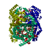
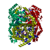
- Structure visualization
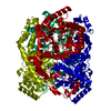
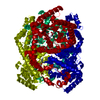
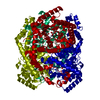
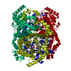
| Structure viewer | Molecule: MolmilJmol/JSmol |
|---|
- Downloads & links
-Download
| PDBx/mmCIF format | 1oad.cif.gz | 189.3 KB | Display | PDBx/mmCIF format |
|---|---|---|---|---|
| PDB format | pdb1oad.ent.gz | 148.2 KB | Display | PDB format |
| PDBx/mmJSON format | 1oad.json.gz | Tree view | PDBx/mmJSON format | |
| Others |  Other downloads Other downloads |
-Validation report
| Arichive directory | https://data.pdbj.org/pub/pdb/validation_reports/oa/1oadftp://data.pdbj.org/pub/pdb/validation_reports/oa/1oad | HTTPS FTP |
|---|
-Related structure data
| Related structure data | |
|---|---|
| Similar structure data |


-Links
 PDBj
PDBj
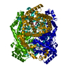
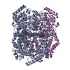
- Assembly
| Deposited unit | 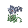
| ||||||||
|---|---|---|---|---|---|---|---|---|---|
| 1 | 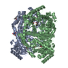
| ||||||||
| Unit cell |
| ||||||||
| Noncrystallographic symmetry (NCS) | NCS oper: (Code: given Matrix: (0.68035, -0.73289, 0.00093), Vector: |
-Components
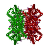
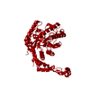
-Protein , 1 types, 2 molecules AB
| #1: Protein | Mass: 43284.285 Da / Num. of mol.: 2 / Source method: isolated from a natural source / Source: (natural) STREPTOMYCES RUBIGINOSUS (bacteria) / References: UniProt: P24300, xylose isomerase |
|---|
-Non-polymers , 7 types, 921 molecules 


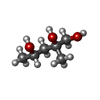


| #2: Chemical |  Mass: 54.938 Da / Num. of mol.: 2 / Source method: obtained synthetically / Formula: Mn Mass: 54.938 Da / Num. of mol.: 2 / Source method: obtained synthetically / Formula: Mn#3: Chemical |  Mass: 24.305 Da / Num. of mol.: 2 / Source method: obtained synthetically / Formula: Mg Mass: 24.305 Da / Num. of mol.: 2 / Source method: obtained synthetically / Formula: Mg#4: Chemical |  Mass: 118.174 Da / Num. of mol.: 2 / Source method: obtained synthetically / Formula: C6H14O2 / Comment: precipitant*YM Mass: 118.174 Da / Num. of mol.: 2 / Source method: obtained synthetically / Formula: C6H14O2 / Comment: precipitant*YM#5: Chemical |  Mass: 134.174 Da / Num. of mol.: 2 / Source method: obtained synthetically / Formula: C6H14O3 Mass: 134.174 Da / Num. of mol.: 2 / Source method: obtained synthetically / Formula: C6H14O3#6: Chemical | ChemComp-TRS / |  Mass: 122.143 Da / Num. of mol.: 1 / Source method: obtained synthetically / Formula: C4H12NO3 / Comment: pH buffer*YM Mass: 122.143 Da / Num. of mol.: 1 / Source method: obtained synthetically / Formula: C4H12NO3 / Comment: pH buffer*YM#7: Chemical | ChemComp-MPD / ( |  Mass: 118.174 Da / Num. of mol.: 1 / Source method: obtained synthetically / Formula: C6H14O2 / Comment: precipitant*YM Mass: 118.174 Da / Num. of mol.: 1 / Source method: obtained synthetically / Formula: C6H14O2 / Comment: precipitant*YM#8: Water | ChemComp-HOH / | Mass: 18.015 Da / Num. of mol.: 911 / Source method: isolated from a natural source / Formula: H2O |
|---|
-Details
| Sequence details | N-TERMINAL MET NOT VISIBLE, NEXT RESIDUE (ASN) WITHOUT SIDE |
|---|
-Experimental details
-Experiment
| Experiment | Method: X-RAY DIFFRACTION / Number of used crystals: 1 |
|---|
- Sample preparation
| Crystal | Density Matthews: 2.89 Å3/Da / Density % sol: 57 % | ||||||||||||||||||||||||||||||||||||||||||
|---|---|---|---|---|---|---|---|---|---|---|---|---|---|---|---|---|---|---|---|---|---|---|---|---|---|---|---|---|---|---|---|---|---|---|---|---|---|---|---|---|---|---|---|
| Crystal grow | pH: 7 Details: 17 MG/ML PROTEIN, 12 % MPD, 0.1 M MGCL2, 50 MM TRIS BUFFER PH 7.0 | ||||||||||||||||||||||||||||||||||||||||||
| Crystal grow | *PLUS Method: vapor diffusion, hanging drop | ||||||||||||||||||||||||||||||||||||||||||
| Components of the solutions | *PLUS
|
-Data collection
| Diffraction | Mean temperature: 100 K |
|---|---|
| Diffraction source | Source: SYNCHROTRON / Site: NSLS  / Beamline: X9B / Wavelength: 1.54 / Beamline: X9B / Wavelength: 1.54 |
| Detector | Type: ADSC CCD / Detector: CCD / Date: Jan 20, 2002 / Details: FOCUSSING MIRROR |
| Radiation | Monochromator: SI (111) DOUBLE CRYSTAL / Protocol: SINGLE WAVELENGTH / Monochromatic (M) / Laue (L): M / Scattering type: x-ray |
| Radiation wavelength | Wavelength: 1.54 Å / Relative weight: 1 |
| Reflection | Resolution: 1.5→35 Å / Num. obs: 300270 / % possible obs: 96.8 % / Observed criterion σ(I): -3 / Redundancy: 7 % / Biso Wilson estimate: 16.3 Å2 / Rmerge(I) obs: 0.056 / Net I/σ(I): 18.8 |
| Reflection shell | Resolution: 1.5→1.55 Å / Redundancy: 3.5 % / Rmerge(I) obs: 0.15 / Mean I/σ(I) obs: 5.6 / % possible all: 93.2 |
| Reflection | *PLUS Lowest resolution: 30 Å / Redundancy: 3.5 % |
| Reflection shell | *PLUS % possible obs: 93.2 % / Rmerge(I) obs: 0.15 |
- Processing
| Software |
| ||||||||||||||||||||
|---|---|---|---|---|---|---|---|---|---|---|---|---|---|---|---|---|---|---|---|---|---|
| Refinement | Method to determine structure: OTHER / Resolution: 1.5→20 Å / Cross valid method: THROUGHOUT / ESU R: 0.06 / ESU R Free: 0.062
| ||||||||||||||||||||
| Displacement parameters | Biso mean: 17.2 Å2 | ||||||||||||||||||||
| Refinement step | Cycle: LAST / Resolution: 1.5→20 Å
| ||||||||||||||||||||
| Refinement | *PLUS Lowest resolution: 19.9 Å | ||||||||||||||||||||
| Solvent computation | *PLUS | ||||||||||||||||||||
| Displacement parameters | *PLUS | ||||||||||||||||||||
| Refine LS restraints | *PLUS
| ||||||||||||||||||||
| LS refinement shell | *PLUS Highest resolution: 1.5 Å / Lowest resolution: 1.573 Å / Rfactor Rfree: 0.29 / Rfactor Rwork: 0.254 / Num. reflection Rwork: 961 / Total num. of bins used: 20 |
