 Movie
Movie Controller

[English] 日本語
 Yorodumi
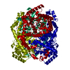
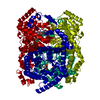
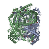
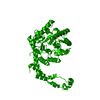
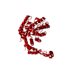
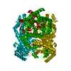
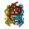
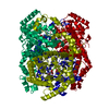
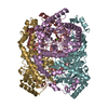
- PDB-9xia: X-RAY ANALYSIS OF D-XYLOSE ISOMERASE AT 1.9 ANGSTROMS: NATIVE ENZ... -
+ Open data

- Basic information
| Entry | Database: PDB / ID: 9xia | |||||||||
|---|---|---|---|---|---|---|---|---|---|---|
| Title | X-RAY ANALYSIS OF D-XYLOSE ISOMERASE AT 1.9 ANGSTROMS: NATIVE ENZYME IN COMPLEX WITH SUBSTRATE AND WITH A MECHANISM-DESIGNED INACTIVATOR | |||||||||
 Components Components | XYLOSE ISOMERASE | |||||||||
 Keywords Keywords | ISOMERASE(INTRAMOLECULAR OXIDOREDUCTASE) | |||||||||
| Function / homology |  Function and homology information Function and homology informationxylose isomerase / xylose isomerase activity / D-xylose metabolic process / magnesium ion binding / identical protein binding / cytoplasm Similarity search - Function | |||||||||
| Biological species |  Streptomyces rubiginosus (bacteria) Streptomyces rubiginosus (bacteria) | |||||||||
| Method |  X-RAY DIFFRACTION / Resolution: 1.9 Å X-RAY DIFFRACTION / Resolution: 1.9 Å | |||||||||
 Authors Authors | Carrell, H.L. / Glusker, J.P. | |||||||||
 Citation Citation | Journal: Proc.Natl.Acad.Sci.USA / Year: 1989 Title: X-ray analysis of D-xylose isomerase at 1.9 A: native enzyme in complex with substrate and with a mechanism-designed inactivator. Authors: Carrell, H.L. / Glusker, J.P. / Burger, V. / Manfre, F. / Tritsch, D. / Biellmann, J.F. #1: Journal: Protein Eng. / Year: 1987Title: Comparison of Backbone Structures of Glucose Isomerase from Streptomyces and Arthrobacter Authors: Henrick, K. / Blow, D.M. / Carrell, H.L. / Glusker, J.P. #2: Journal: J.Biol.Chem. / Year: 1984Title: X-Ray Crystal Structure of D-Xylose Isomerase at 4-Angstroms Resolution Authors: Carrell, H.L. / Rubin, B.H. / Hurley, T.J. / Glusker, J.P. | |||||||||
| History |
| |||||||||
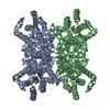
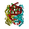
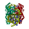
| Remark 700 | SHEET THE STRUCTURE OF THE MONOMER IS AN EIGHT-FOLD ALPHA-BETA BARREL WITH AN EXTENDED C-TERMINAL ...SHEET THE STRUCTURE OF THE MONOMER IS AN EIGHT-FOLD ALPHA-BETA BARREL WITH AN EXTENDED C-TERMINAL LOOP WHICH FACILITATES AGGREGATION OF MONOMERS TO TETRAMERS. TETRAMERS ARE POSITIONED ON THE 222 SYMMETRY SITE AT THE ORIGIN OF THE CELL. |
- Structure visualization
| Structure viewer | Molecule: MolmilJmol/JSmol |
|---|
- Downloads & links
-Download
| PDBx/mmCIF format | 9xia.cif.gz | 95.4 KB | Display | PDBx/mmCIF format |
|---|---|---|---|---|
| PDB format | pdb9xia.ent.gz | 71.3 KB | Display | PDB format |
| PDBx/mmJSON format | 9xia.json.gz | Tree view | PDBx/mmJSON format | |
| Others |  Other downloads Other downloads |
-Validation report
| Arichive directory | https://data.pdbj.org/pub/pdb/validation_reports/xi/9xiaftp://data.pdbj.org/pub/pdb/validation_reports/xi/9xia | HTTPS FTP |
|---|
-Related structure data


-Links
 PDBj
PDBj
- Assembly
| Deposited unit | 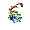
| |||||||||
|---|---|---|---|---|---|---|---|---|---|---|
| 1 | 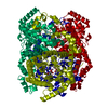
| |||||||||
| Unit cell |
| |||||||||
| Atom site foot note | 1: RESIDUE PRO 187 IS A CIS PROLINE. | |||||||||
| Components on special symmetry positions |
| |||||||||
| Details | THE STRUCTURE OF THE MONOMER IS AN EIGHT-FOLD ALPHA-BETA BARREL WITH AN EXTENDED C-TERMINAL LOOP WHICH FACILITATES AGGREGATION OF MONOMERS TO TETRAMERS. TETRAMERS ARE POSITIONED ON THE 222 SYMMETRY SITE AT THE ORIGIN OF THE CELL. |
-Components
| #1: Protein | Mass: 43254.234 Da / Num. of mol.: 1 Source method: isolated from a genetically manipulated source Source: (gene. exp.) Streptomyces rubiginosus (bacteria) / References: UniProt: P24300, xylose isomerase | ||||||
|---|---|---|---|---|---|---|---|
| #2: Sugar | ChemComp-DFR / 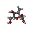  Type: D-saccharide, beta linking / Mass: 178.183 Da / Num. of mol.: 1 Type: D-saccharide, beta linking / Mass: 178.183 Da / Num. of mol.: 1Source method: isolated from a genetically manipulated source Formula: C7H14O5 | ||||||
| #3: Chemical |   Mass: 54.938 Da / Num. of mol.: 2 / Source method: obtained synthetically / Formula: Mn Mass: 54.938 Da / Num. of mol.: 2 / Source method: obtained synthetically / Formula: Mn#4: Water | ChemComp-HOH / |  Mass: 18.015 Da / Num. of mol.: 299 / Source method: isolated from a natural source / Formula: H2O Mass: 18.015 Da / Num. of mol.: 299 / Source method: isolated from a natural source / Formula: H2OHas protein modification | Y | Sequence details | THE AMINO ACID SEQUENCE WAS TAKEN FROM INTERNATIONAL PATENT APPLICATION NUMBER:PCT/US88/02765, ...THE AMINO ACID SEQUENCE WAS TAKEN FROM INTERNATIO | |
-Experimental details
-Experiment
| Experiment | Method: X-RAY DIFFRACTION |
|---|
- Sample preparation
| Crystal | Density Matthews: 2.79 Å3/Da / Density % sol: 55.93 % | ||||||||||||||||||||
|---|---|---|---|---|---|---|---|---|---|---|---|---|---|---|---|---|---|---|---|---|---|
| Crystal grow | *PLUS Temperature: 4 ℃ / pH: 7.4 / Method: batch method | ||||||||||||||||||||
| Components of the solutions | *PLUS
|
-Data collection
| Radiation | Scattering type: x-ray |
|---|---|
| Radiation wavelength | Relative weight: 1 |
| Reflection | *PLUS Highest resolution: 1.9 Å / Num. obs: 36274 / % possible obs: 94 % / Num. measured all: 71368 / Biso Wilson estimate: 22 Å2 |
- Processing
| Software | Name: PROLSQ / Classification: refinement | |||||||||||||||||||||||||||||||||||||||||||||||||||||||||||||||
|---|---|---|---|---|---|---|---|---|---|---|---|---|---|---|---|---|---|---|---|---|---|---|---|---|---|---|---|---|---|---|---|---|---|---|---|---|---|---|---|---|---|---|---|---|---|---|---|---|---|---|---|---|---|---|---|---|---|---|---|---|---|---|---|---|
| Refinement | Resolution: 1.9→8 Å /
| |||||||||||||||||||||||||||||||||||||||||||||||||||||||||||||||
| Refinement step | Cycle: LAST / Resolution: 1.9→8 Å
| |||||||||||||||||||||||||||||||||||||||||||||||||||||||||||||||
| Refine LS restraints |
| |||||||||||||||||||||||||||||||||||||||||||||||||||||||||||||||
| Software | *PLUS Name: PROLSQ / Classification: refinement | |||||||||||||||||||||||||||||||||||||||||||||||||||||||||||||||
| Refinement | *PLUS Highest resolution: 1.9 Å / Lowest resolution: 8 Å / Num. reflection obs: 30250 / σ(I): 1.5 | |||||||||||||||||||||||||||||||||||||||||||||||||||||||||||||||
| Solvent computation | *PLUS | |||||||||||||||||||||||||||||||||||||||||||||||||||||||||||||||
| Displacement parameters | *PLUS Biso mean: 17 Å2 |
