[English] 日本語
 Yorodumi
- PDB-1xya: X-RAY CRYSTALLOGRAPHIC STRUCTURES OF D-XYLOSE ISOMERASE-SUBSTRATE... -
+ Open data
- Basic information
| Entry | Database: PDB / ID: 1xya | |||||||||
|---|---|---|---|---|---|---|---|---|---|---|
| Title | X-RAY CRYSTALLOGRAPHIC STRUCTURES OF D-XYLOSE ISOMERASE-SUBSTRATE COMPLEXES POSITION THE SUBSTRATE AND PROVIDE EVIDENCE FOR METAL MOVEMENT DURING CATALYSIS | |||||||||
 Components Components | XYLOSE ISOMERASE | |||||||||
 Keywords Keywords | ISOMERASE / OXIDOREDUCTASE / ISOMERASE(INTRAMOLECULAR OXIDOREDUCTASE) | |||||||||
| Function / homology |  Function and homology information Function and homology informationxylose isomerase / xylose isomerase activity / D-xylose metabolic process / magnesium ion binding / cytoplasm Similarity search - Function | |||||||||
| Biological species |  Streptomyces olivochromogenes (bacteria) Streptomyces olivochromogenes (bacteria) | |||||||||
| Method |  X-RAY DIFFRACTION / Resolution: 1.81 Å X-RAY DIFFRACTION / Resolution: 1.81 Å | |||||||||
 Authors Authors | Lavie, A. / Allen, K.N. / Petsko, G.A. / Ringe, D. | |||||||||
 Citation Citation |  Journal: Biochemistry / Year: 1994 Journal: Biochemistry / Year: 1994Title: X-ray crystallographic structures of D-xylose isomerase-substrate complexes position the substrate and provide evidence for metal movement during catalysis. Authors: Lavie, A. / Allen, K.N. / Petsko, G.A. / Ringe, D. #1:  Journal: Biochemistry / Year: 1994 Journal: Biochemistry / Year: 1994Title: Isotopic Exchange Plus Substrate and Inhibition Kinetics of D-Xylose Isomerase Do not Support a Proton-Transfer Mechanism Authors: Allen, K.N. / Lavie, A. / Farber, G.K. / Glasfeld, A. / Petsko, G.A. / Ringe, D. #2:  Journal: Biochemistry / Year: 1994 Journal: Biochemistry / Year: 1994Title: The Role of the Divalent Metal Ion in Sugar Binding, Ring Opening, and Isomerization by D-Xylose Isomerase: Replacement of a Catalytic Metal by an Amino-Acid Authors: Allen, K.N. / Lavie, A. / Glasfeld, A. / Tanada, T.N. / Gerrity, D.P. / Carlson, S.C. / Farber, G.K. / Petsko, G.A. / Ringe, D. | |||||||||
| History |
| |||||||||
| Remark 700 | SHEET THE SHEETS PRESENTED AS *SA1*, *SA2*, *SB1*, AND *SB2* ON SHEET RECORDS BELOW ARE ACTUALLY ...SHEET THE SHEETS PRESENTED AS *SA1*, *SA2*, *SB1*, AND *SB2* ON SHEET RECORDS BELOW ARE ACTUALLY EIGHT-STRANDED BETA-BARRELS. THESE ARE REPRESENTED AS NINE-STRANDED SHEETS IN WHICH THE FIRST AND LAST STRANDS OF EACH SHEET ARE IDENTICAL. THERE ARE SEVERAL BIFURCATED SHEETS IN THIS STRUCTURE. THESE ARE REPRESENTED BY TWO SHEETS WHICH HAVE ONE OR MORE IDENTICAL STRANDS. SHEETS *SA1* AND *SB1* REPRESENT ONE BIFURCATED SHEET. SHEETS *SA2* AND *SB2* REPRESENT ANOTHER BIFURCATED SHEET. |
- Structure visualization
| Structure viewer | Molecule:  Molmil Molmil Jmol/JSmol Jmol/JSmol |
|---|
- Downloads & links
- Download
| PDBx/mmCIF format |  1xya.cif.gz 1xya.cif.gz | 172.5 KB | Display |  PDBx/mmCIF format PDBx/mmCIF format |
|---|---|---|---|---|
| PDB format |  pdb1xya.ent.gz pdb1xya.ent.gz | 134.7 KB | Display |  PDB format PDB format |
| PDBx/mmJSON format |  1xya.json.gz 1xya.json.gz | Tree view |  PDBx/mmJSON format PDBx/mmJSON format | |
| Others |  Other downloads Other downloads |
-Validation report
| Arichive directory |  https://data.pdbj.org/pub/pdb/validation_reports/xy/1xya https://data.pdbj.org/pub/pdb/validation_reports/xy/1xya ftp://data.pdbj.org/pub/pdb/validation_reports/xy/1xya ftp://data.pdbj.org/pub/pdb/validation_reports/xy/1xya | HTTPS FTP |
|---|
-Related structure data
- Links
Links
- Assembly
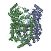
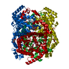
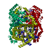
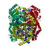
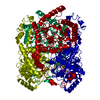
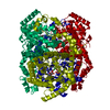
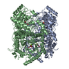
| Deposited unit | 
| ||||||||
|---|---|---|---|---|---|---|---|---|---|
| 1 | 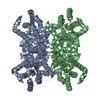
| ||||||||
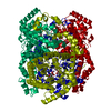
| Unit cell |
| ||||||||
| Atom site foot note | 1: CIS PROLINE - PRO A 186 / 2: CIS PROLINE - PRO B 686 | ||||||||
| Noncrystallographic symmetry (NCS) | NCS oper: (Code: given Matrix: (0.99927, 0.03829, -0.00045), Vector: |
- Components
| #1: Protein | Mass: 42844.848 Da / Num. of mol.: 2 Source method: isolated from a genetically manipulated source Source: (gene. exp.)  Streptomyces olivochromogenes (bacteria) Streptomyces olivochromogenes (bacteria)References: UniProt: P15587, xylose isomerase #2: Chemical | ChemComp-MG / #3: Chemical | #4: Water | ChemComp-HOH / | Sequence details | THE SEQUENCE REPORTED HERE DISAGREES WITH THAT ORIGINALLY REPORTED (FARBER ET AL., BIOCHEMISTRY V. ...THE SEQUENCE REPORTED HERE DISAGREES WITH THAT ORIGINALLY | |
|---|
-Experimental details
-Experiment
| Experiment | Method:  X-RAY DIFFRACTION X-RAY DIFFRACTION |
|---|
- Sample preparation
| Crystal | Density Matthews: 2.39 Å3/Da / Density % sol: 48.53 % | ||||||||||||||||||||||||||||||||||||
|---|---|---|---|---|---|---|---|---|---|---|---|---|---|---|---|---|---|---|---|---|---|---|---|---|---|---|---|---|---|---|---|---|---|---|---|---|---|
| Crystal grow | *PLUS Method: vapor diffusion, sitting dropDetails: taken from Farber, G.K. et al (1987). Protein Eng., 1, 459-466. pH: 7.5 | ||||||||||||||||||||||||||||||||||||
| Components of the solutions | *PLUS
|
-Data collection
| Reflection | *PLUS Highest resolution: 1.81 Å / Lowest resolution: 9999 Å / Num. obs: 56756 / % possible obs: 75 % / Num. measured all: 243152 / Rmerge(I) obs: 0.051 |
|---|
- Processing
| Software |
| ||||||||||||||||||||||||||||||||||||||||||||||||||||||||||||
|---|---|---|---|---|---|---|---|---|---|---|---|---|---|---|---|---|---|---|---|---|---|---|---|---|---|---|---|---|---|---|---|---|---|---|---|---|---|---|---|---|---|---|---|---|---|---|---|---|---|---|---|---|---|---|---|---|---|---|---|---|---|
| Refinement | Resolution: 1.81→10 Å / Rfactor Rwork: 0.161 / Rfactor obs: 0.161 | ||||||||||||||||||||||||||||||||||||||||||||||||||||||||||||
| Refinement step | Cycle: LAST / Resolution: 1.81→10 Å
| ||||||||||||||||||||||||||||||||||||||||||||||||||||||||||||
| Refine LS restraints |
| ||||||||||||||||||||||||||||||||||||||||||||||||||||||||||||
| Refinement | *PLUS Rfactor obs: 0.161 / Rfactor Rwork: 0.161 | ||||||||||||||||||||||||||||||||||||||||||||||||||||||||||||
| Solvent computation | *PLUS | ||||||||||||||||||||||||||||||||||||||||||||||||||||||||||||
| Displacement parameters | *PLUS | ||||||||||||||||||||||||||||||||||||||||||||||||||||||||||||
| Refine LS restraints | *PLUS
|
 Movie
Movie Controller


 PDBj
PDBj


