 Movie
Movie Controller
[English] 日本語
 Yorodumi
- SASDCK2: Glucose Isomerase - Streptomyces rubiginosus (Xylose isomerase, G... -
+ Open data

- Basic information
| Entry | Database: SASBDB / ID: SASDCK2 |
|---|---|
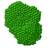
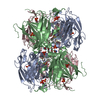
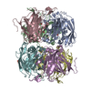
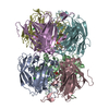
 Sample Sample | Glucose Isomerase - Streptomyces rubiginosus
|
| Function / homology |  Function and homology information Function and homology informationxylose isomerase / xylose isomerase activity / D-xylose metabolic process / magnesium ion binding / identical protein binding / cytoplasm Similarity search - Function |
| Biological species |  Streptomyces rubiginosus (bacteria) Streptomyces rubiginosus (bacteria) |
 Citation Citation | Journal: Acta Crystallogr D Struct Biol / Year: 2017 Title: 2017 publication guidelines for structural modelling of small-angle scattering data from biomolecules in solution: an update. Authors: Jill Trewhella / Anthony P Duff / Dominique Durand / Frank Gabel / J Mitchell Guss / Wayne A Hendrickson / Greg L Hura / David A Jacques / Nigel M Kirby / Ann H Kwan / Javier Pérez / Lois ...Authors: Jill Trewhella / Anthony P Duff / Dominique Durand / Frank Gabel / J Mitchell Guss / Wayne A Hendrickson / Greg L Hura / David A Jacques / Nigel M Kirby / Ann H Kwan / Javier Pérez / Lois Pollack / Timothy M Ryan / Andrej Sali / Dina Schneidman-Duhovny / Torsten Schwede / Dmitri I Svergun / Masaaki Sugiyama / John A Tainer / Patrice Vachette / John Westbrook / Andrew E Whitten /        Abstract: In 2012, preliminary guidelines were published addressing sample quality, data acquisition and reduction, presentation of scattering data and validation, and modelling for biomolecular small-angle ...In 2012, preliminary guidelines were published addressing sample quality, data acquisition and reduction, presentation of scattering data and validation, and modelling for biomolecular small-angle scattering (SAS) experiments. Biomolecular SAS has since continued to grow and authors have increasingly adopted the preliminary guidelines. In parallel, integrative/hybrid determination of biomolecular structures is a rapidly growing field that is expanding the scope of structural biology. For SAS to contribute maximally to this field, it is essential to ensure open access to the information required for evaluation of the quality of SAS samples and data, as well as the validity of SAS-based structural models. To this end, the preliminary guidelines for data presentation in a publication are reviewed and updated, and the deposition of data and associated models in a public archive is recommended. These guidelines and recommendations have been prepared in consultation with the members of the International Union of Crystallography (IUCr) Small-Angle Scattering and Journals Commissions, the Worldwide Protein Data Bank (wwPDB) Small-Angle Scattering Validation Task Force and additional experts in the field. |
 Contact author Contact author |
|
- Structure visualization
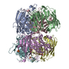
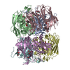
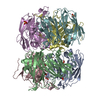
| Structure viewer | Molecule: MolmilJmol/JSmol |
|---|
- Downloads & links
-Data source
| SASBDB page |  SASDCK2 SASDCK2 |
|---|
-Related structure data
| Related structure data | C: citing same article ( |
|---|---|
| Similar structure data |


-External links
| Related items in Molecule of the Month |
|---|

-Models
| Model #1173 |   Type: dummy / Software: (on-line) / Radius of dummy atoms: 2.40 A / Symmetry: P1 / Comment: job submitted ATSAS on-line April 9, 2017 / Chi-square value: 0.951 / P-value: 0.035000  Search similar-shape structures of this assembly by Omokage search (details) Search similar-shape structures of this assembly by Omokage search (details) |
|---|---|
| Model #1174 |  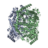 Type: atomic / Software: (on-line) / Radius of dummy atoms: 1.90 A Comment: GI tetramer generated from PDB:1OAD using symmetry transforms in pymol Chi-square value: 1.01580033274 / P-value: 0.045511 Search similar-shape structures of this assembly by Omokage search (details) |
-Sample
| Sample | Name: Glucose Isomerase - Streptomyces rubiginosus / Specimen concentration: 0.06-0.34 |
|---|---|
| Buffer | Name: 25 mM MOPS, 250 mM NaCl, 50 mM KCl, 2 mM TCEP, 0.1% NaN3 pH: 7.5 Comment: 2 mM TCEP and 0.1% (.01538 M) NaN3 used for radiation protection |
| Entity #612 | Name: Glucose Isomerase / Type: protein / Description: Xylose isomerase / Formula weight: 43.097 / Num. of mol.: 4 / Source: Streptomyces rubiginosus / References: UniProt: P24300 Sequence: NYQPTPEDRF TFGLWTVGWE GRDPFGDATR RALDPVESVR RLAELGAHGV TFHDDDLIPF GSSDSEREEH VKRFRQALDD TGMKVPMATT NLFTHPVFKD GGFTANDRDV RRYALRKTIR NIDLAVELGA ETYVAWGGRE GAESGGAKDV RDALDRMKEA FDLLGEYVTS ...Sequence: NYQPTPEDRF TFGLWTVGWE GRDPFGDATR RALDPVESVR RLAELGAHGV TFHDDDLIPF GSSDSEREEH VKRFRQALDD TGMKVPMATT NLFTHPVFKD GGFTANDRDV RRYALRKTIR NIDLAVELGA ETYVAWGGRE GAESGGAKDV RDALDRMKEA FDLLGEYVTS QGYDIRFAIE PKPNEPRGDI LLPTVGHALA FIERLERPEL YGVNPEVGHE QMAGLNFPHG IAQALWAGKL FHIDLNGQNG IKYDQDLRFG AGDLRAAFWL VDLLESAGYS GPRHFDFKPP RTEDFDGVWA SAAGCMRNYL ILKERAAAFR ADPEVQEALR ASRLDELARP TAADGLQALL DDRSAFEEFD VDAAAARGMA FERLDQLAMD HLLGARG |
-Experimental information
| Beam | Instrument name: Australian Synchrotron SAXS/WAXS / City: Melbourne / 国: Australia / Shape: Point / Type of source: X-ray synchrotron / Wavelength: 0.10332 Å / Dist. spec. to detc.: 2.683 mm | ||||||||||||||||||||||||||||||||||||
|---|---|---|---|---|---|---|---|---|---|---|---|---|---|---|---|---|---|---|---|---|---|---|---|---|---|---|---|---|---|---|---|---|---|---|---|---|---|
| Detector | Name: Pilatus 1M / Type: Dectris / Pixsize x: 172 mm | ||||||||||||||||||||||||||||||||||||
| Scan | 
| ||||||||||||||||||||||||||||||||||||
| Distance distribution function P(R) | 
| ||||||||||||||||||||||||||||||||||||
| Result | 
|
