 Movie
Movie Controller

[English] 日本語
 Yorodumi
- PDB-4zb0: A dehydrated form of glucose isomerase collected at room temperature. -
+ Open data

- Basic information
| Entry | Database: PDB / ID: 4zb0 | ||||||
|---|---|---|---|---|---|---|---|
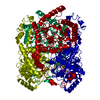
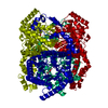
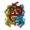
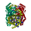
| Title | A dehydrated form of glucose isomerase collected at room temperature. | ||||||
 Components Components | Xylose isomerase | ||||||
 Keywords Keywords | ISOMERASE | ||||||
| Function / homology |  Function and homology information Function and homology informationxylose isomerase / xylose isomerase activity / D-xylose metabolic process / magnesium ion binding / identical protein binding / cytoplasm Similarity search - Function | ||||||
| Biological species |  Streptomyces rubiginosus (bacteria) Streptomyces rubiginosus (bacteria) | ||||||
| Method |  X-RAY DIFFRACTION / SYNCHROTRON / MOLECULAR REPLACEMENT / Resolution: 2 Å X-RAY DIFFRACTION / SYNCHROTRON / MOLECULAR REPLACEMENT / Resolution: 2 Å | ||||||
 Authors Authors | Sandy, J. | ||||||
 Citation Citation | Journal: Acta Crystallogr D Struct Biol / Year: 2016 Title: A generic protocol for protein crystal dehydration using the HC1b humidity controller. Authors: Lobley, C.M. / Sandy, J. / Sanchez-Weatherby, J. / Mazzorana, M. / Krojer, T. / Nowak, R.P. / Sorensen, T.L. | ||||||
| History |
|
- Structure visualization
| Structure viewer | Molecule: MolmilJmol/JSmol |
|---|
- Downloads & links
-Download
| PDBx/mmCIF format | 4zb0.cif.gz | 174.2 KB | Display | PDBx/mmCIF format |
|---|---|---|---|---|
| PDB format | pdb4zb0.ent.gz | 136.2 KB | Display | PDB format |
| PDBx/mmJSON format | 4zb0.json.gz | Tree view | PDBx/mmJSON format | |
| Others |  Other downloads Other downloads |
-Validation report
| Arichive directory | https://data.pdbj.org/pub/pdb/validation_reports/zb/4zb0ftp://data.pdbj.org/pub/pdb/validation_reports/zb/4zb0 | HTTPS FTP |
|---|
-Related structure data
| Related structure data |  4zb2C  4zb5C  4zbcC  1mnzS C: citing same article ( S: Starting model for refinement |
|---|---|
| Similar structure data |


-Links
 PDBj
PDBj


- Assembly
| Deposited unit | 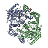
| ||||||||
|---|---|---|---|---|---|---|---|---|---|
| 1 | 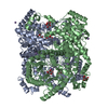
| ||||||||
| Unit cell |
|
-Components
| #1: Protein | Mass: 43152.105 Da / Num. of mol.: 2 / Fragment: UNP residues 2-388 Source method: isolated from a genetically manipulated source Source: (gene. exp.) Streptomyces rubiginosus (bacteria) / Gene: xylA / Production host: Streptomyces rubiginosus (bacteria) / References: UniProt: P24300, xylose isomerase#2: Chemical | ChemComp-MN /   Mass: 54.938 Da / Num. of mol.: 10 / Source method: obtained synthetically / Formula: Mn Mass: 54.938 Da / Num. of mol.: 10 / Source method: obtained synthetically / Formula: Mn#3: Sugar |   Type: D-saccharide, alpha linking / Mass: 180.156 Da / Num. of mol.: 2 Type: D-saccharide, alpha linking / Mass: 180.156 Da / Num. of mol.: 2Source method: isolated from a genetically manipulated source Formula: C6H12O6 #4: Sugar |   Type: D-saccharide, beta linking / Mass: 180.156 Da / Num. of mol.: 2 Type: D-saccharide, beta linking / Mass: 180.156 Da / Num. of mol.: 2Source method: isolated from a genetically manipulated source Formula: C6H12O6 #5: Water | ChemComp-HOH / |  Mass: 18.015 Da / Num. of mol.: 446 / Source method: isolated from a natural source / Formula: H2O Mass: 18.015 Da / Num. of mol.: 446 / Source method: isolated from a natural source / Formula: H2O |
|---|
-Experimental details
-Experiment
| Experiment | Method: X-RAY DIFFRACTION |
|---|
- Sample preparation
| Crystal | Density Matthews: 2.26 Å3/Da / Density % sol: 45.65 % |
|---|---|
| Crystal grow | Temperature: 293 K / Method: vapor diffusion, sitting drop / pH: 7 Details: 0.1 M Hepes, 10% PEG 400, 0.05 M Manganese Chloride, 20% Glucose |
-Data collection
| Diffraction | Mean temperature: 293 K Ambient temp details: Collected using HC1b humidity control device at RH of 96% |
|---|---|
| Diffraction source | Source: SYNCHROTRON / Site: Diamond  / Beamline: I02 / Wavelength: 0.9795 Å / Beamline: I02 / Wavelength: 0.9795 Å |
| Detector | Type: DECTRIS PILATUS 6M / Detector: PIXEL / Date: Feb 9, 2015 |
| Radiation | Monochromator: Si (111) / Protocol: SINGLE WAVELENGTH / Monochromatic (M) / Laue (L): M / Scattering type: x-ray |
| Radiation wavelength | Wavelength: 0.9795 Å / Relative weight: 1 |
| Reflection | Resolution: 2→68.38 Å / Num. obs: 53324 / % possible obs: 99.5 % / Redundancy: 3.2 % / Rmerge(I) obs: 0.06 / Net I/σ(I): 10.4 / Num. measured all: 169888 |
| Reflection shell | Resolution: 2→2.1 Å / Redundancy: 3.3 % / Rmerge(I) obs: 0.138 / Mean I/σ(I) obs: 5.9 / % possible all: 99.6 |
- Processing
| Software |
| ||||||||||||||||||||||||||||||||||||||||||||||||||||||||||||||||||||||||||||||||||||||||||||||||||||||||||||||||||||||||||||||||||||||||||||||||||||||||||||||||||||||||||||||||||||||
|---|---|---|---|---|---|---|---|---|---|---|---|---|---|---|---|---|---|---|---|---|---|---|---|---|---|---|---|---|---|---|---|---|---|---|---|---|---|---|---|---|---|---|---|---|---|---|---|---|---|---|---|---|---|---|---|---|---|---|---|---|---|---|---|---|---|---|---|---|---|---|---|---|---|---|---|---|---|---|---|---|---|---|---|---|---|---|---|---|---|---|---|---|---|---|---|---|---|---|---|---|---|---|---|---|---|---|---|---|---|---|---|---|---|---|---|---|---|---|---|---|---|---|---|---|---|---|---|---|---|---|---|---|---|---|---|---|---|---|---|---|---|---|---|---|---|---|---|---|---|---|---|---|---|---|---|---|---|---|---|---|---|---|---|---|---|---|---|---|---|---|---|---|---|---|---|---|---|---|---|---|---|---|---|
| Refinement | Method to determine structure: MOLECULAR REPLACEMENT Starting model: 1MNZ Resolution: 2→68.38 Å / Cor.coef. Fo:Fc: 0.967 / Cor.coef. Fo:Fc free: 0.948 / SU B: 3.183 / SU ML: 0.088 / Cross valid method: THROUGHOUT / ESU R: 0.146 / ESU R Free: 0.132 / Stereochemistry target values: MAXIMUM LIKELIHOOD / Details: HYDROGENS HAVE BEEN ADDED IN THE RIDING POSITIONS
| ||||||||||||||||||||||||||||||||||||||||||||||||||||||||||||||||||||||||||||||||||||||||||||||||||||||||||||||||||||||||||||||||||||||||||||||||||||||||||||||||||||||||||||||||||||||
| Solvent computation | Ion probe radii: 0.8 Å / Shrinkage radii: 0.8 Å / VDW probe radii: 1.2 Å / Solvent model: MASK | ||||||||||||||||||||||||||||||||||||||||||||||||||||||||||||||||||||||||||||||||||||||||||||||||||||||||||||||||||||||||||||||||||||||||||||||||||||||||||||||||||||||||||||||||||||||
| Displacement parameters | Biso mean: 20.946 Å2
| ||||||||||||||||||||||||||||||||||||||||||||||||||||||||||||||||||||||||||||||||||||||||||||||||||||||||||||||||||||||||||||||||||||||||||||||||||||||||||||||||||||||||||||||||||||||
| Refinement step | Cycle: LAST / Resolution: 2→68.38 Å
| ||||||||||||||||||||||||||||||||||||||||||||||||||||||||||||||||||||||||||||||||||||||||||||||||||||||||||||||||||||||||||||||||||||||||||||||||||||||||||||||||||||||||||||||||||||||
| Refine LS restraints |
|