 Movie
Movie Controller


+ Open data

- Basic information
| Entry | Database: PDB / ID: 3k51 | ||||||
|---|---|---|---|---|---|---|---|
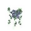
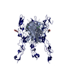
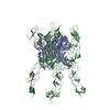
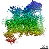
| Title | Crystal Structure of DcR3-TL1A complex | ||||||
 Components Components |
| ||||||
 Keywords Keywords | IMMUNE SYSTEM / DcR3 / TL1A / TNF / TNFR / DECOY RECEPTOR / IMMUNITY / Cytokine / Disulfide bond / Glycoprotein / Membrane / Secreted / Signal-anchor / Transmembrane / Apoptosis / Receptor | ||||||
| Function / homology |  Function and homology information Function and homology informationTNFs bind their physiological receptors / activation of NF-kappaB-inducing kinase activity / tumor necrosis factor receptor binding / positive regulation of extrinsic apoptotic signaling pathway / cytokine activity / signaling receptor activity / positive regulation of canonical NF-kappaB signal transduction / cell surface receptor signaling pathway / immune response / signaling receptor binding ...TNFs bind their physiological receptors / activation of NF-kappaB-inducing kinase activity / tumor necrosis factor receptor binding / positive regulation of extrinsic apoptotic signaling pathway / cytokine activity / signaling receptor activity / positive regulation of canonical NF-kappaB signal transduction / cell surface receptor signaling pathway / immune response / signaling receptor binding / apoptotic process / negative regulation of apoptotic process / signal transduction / : / extracellular region / membrane / plasma membrane Similarity search - Function | ||||||
| Biological species |  Homo sapiens (human) Homo sapiens (human) | ||||||
| Method |  X-RAY DIFFRACTION / SYNCHROTRON / MOLECULAR REPLACEMENT / molecular replacement / Resolution: 2.45 Å X-RAY DIFFRACTION / SYNCHROTRON / MOLECULAR REPLACEMENT / molecular replacement / Resolution: 2.45 Å | ||||||
 Authors Authors | Zhan, C. / Patskovsky, Y. / Yan, Q. / Li, Z. / Ramagopal, U.A. / Nathenson, S.G. / Almo, S.C. | ||||||
 Citation Citation | Journal: Structure / Year: 2011 Title: Decoy Strategies: The Structure of TL1A:DcR3 Complex. Authors: Zhan, C. / Patskovsky, Y. / Yan, Q. / Li, Z. / Ramagopal, U. / Cheng, H. / Brenowitz, M. / Hui, X. / Nathenson, S.G. / Almo, S.C. | ||||||
| History |
|
- Structure visualization
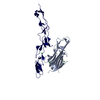
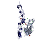
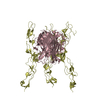
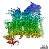
| Structure viewer | Molecule: MolmilJmol/JSmol |
|---|
- Downloads & links
-Download
| PDBx/mmCIF format | 3k51.cif.gz | 76.7 KB | Display | PDBx/mmCIF format |
|---|---|---|---|---|
| PDB format | pdb3k51.ent.gz | 56.5 KB | Display | PDB format |
| PDBx/mmJSON format | 3k51.json.gz | Tree view | PDBx/mmJSON format | |
| Others |  Other downloads Other downloads |
-Validation report
| Arichive directory | https://data.pdbj.org/pub/pdb/validation_reports/k5/3k51ftp://data.pdbj.org/pub/pdb/validation_reports/k5/3k51 | HTTPS FTP |
|---|
-Related structure data
| Related structure data | 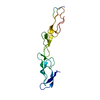 3mhdC  3mi8C  2qe3S S: Starting model for refinement C: citing same article ( |
|---|---|
| Similar structure data |
-Links
 PDBj
PDBj
- Assembly
| Deposited unit | 
| ||||||||
|---|---|---|---|---|---|---|---|---|---|
| 1 | 
| ||||||||
| Unit cell |
|
-Components
| #1: Protein | Mass: 20877.619 Da / Num. of mol.: 1 / Mutation: C95S,C135S Source method: isolated from a genetically manipulated source Source: (gene. exp.) Homo sapiens (human) / Gene: TL1, TNFSF15, VEGI / Plasmid: pET28a / Production host:  |
|---|---|
| #2: Protein | Mass: 19288.488 Da / Num. of mol.: 1 / Fragment: TNFR cysteine rich domain Source method: isolated from a genetically manipulated source Source: (gene. exp.) Homo sapiens (human) / Gene: DCR3, TNFRSF6B, TR6, UNQ186/PRO212 / Plasmid: pMT/BiP/V5-His / Cell line (production host): S2 / Production host:  |
| #3: Water | ChemComp-HOH /  Mass: 18.015 Da / Num. of mol.: 83 / Source method: isolated from a natural source / Formula: H2O Mass: 18.015 Da / Num. of mol.: 83 / Source method: isolated from a natural source / Formula: H2O |
| Has protein modification | Y |
-Experimental details
-Experiment
| Experiment | Method: X-RAY DIFFRACTION / Number of used crystals: 1 |
|---|
- Sample preparation
| Crystal | Density Matthews: 2.88 Å3/Da / Density % sol: 57.36 % |
|---|---|
| Crystal grow | Temperature: 290 K / pH: 6.5 Details: 0.1M MES pH6.5, 8% PEG 10K, 3% dextran sulfate, VAPOR DIFFUSION, SITTING DROP, temperature 290.0K |
-Data collection
| Diffraction | Mean temperature: 100 K |
|---|---|
| Diffraction source | Source: SYNCHROTRON / Site: APS  / Beamline: 24-ID-E / Wavelength: 0.9792 / Beamline: 24-ID-E / Wavelength: 0.9792 |
| Detector | Type: ADSC QUANTUM 315 / Detector: CCD / Date: Nov 8, 2008 |
| Radiation | Protocol: SINGLE WAVELENGTH / Monochromatic (M) / Laue (L): M / Scattering type: x-ray |
| Radiation wavelength | Wavelength: 0.9792 Å / Relative weight: 1 |
| Reflection | Resolution: 2.45→50 Å / Num. obs: 17791 / % possible obs: 99.9 % / Redundancy: 7.7 % / Biso Wilson estimate: 47.85 Å2 / Rmerge(I) obs: 0.131 / Net I/σ(I): 15.3 |
| Reflection shell | Resolution: 2.45→2.49 Å / Redundancy: 7.6 % / Rmerge(I) obs: 0.99 / Mean I/σ(I) obs: 3.17 / % possible all: 100 |
-Phasing
| Phasing | Method: molecular replacement |
|---|
- Processing
| Software |
| ||||||||||||||||||||||||||||||||||||||||||||||||||||||||||||||||||||||||||||||||||||||||||||||||||||||||||||||||||||||||||||||||||||||||||||||||||||||||||||||||||||||||||
|---|---|---|---|---|---|---|---|---|---|---|---|---|---|---|---|---|---|---|---|---|---|---|---|---|---|---|---|---|---|---|---|---|---|---|---|---|---|---|---|---|---|---|---|---|---|---|---|---|---|---|---|---|---|---|---|---|---|---|---|---|---|---|---|---|---|---|---|---|---|---|---|---|---|---|---|---|---|---|---|---|---|---|---|---|---|---|---|---|---|---|---|---|---|---|---|---|---|---|---|---|---|---|---|---|---|---|---|---|---|---|---|---|---|---|---|---|---|---|---|---|---|---|---|---|---|---|---|---|---|---|---|---|---|---|---|---|---|---|---|---|---|---|---|---|---|---|---|---|---|---|---|---|---|---|---|---|---|---|---|---|---|---|---|---|---|---|---|---|---|---|---|
| Refinement | Method to determine structure: MOLECULAR REPLACEMENT Starting model: PDB ENTRY 2QE3 Resolution: 2.45→36.23 Å / Cor.coef. Fo:Fc: 0.93 / Cor.coef. Fo:Fc free: 0.905 / Occupancy max: 1 / Occupancy min: 0.33 / SU B: 6.811 / SU ML: 0.163 / Cross valid method: THROUGHOUT / ESU R: 0.324 / ESU R Free: 0.247 / Stereochemistry target values: MAXIMUM LIKELIHOOD Details: EXTRA ELECTRON DENSITY POSSIBLY CORRESPONDING TO N-GLYCAN IS OBSERVED CLOSE TO N173 IN CHAIN B. BECAUSE OF THE DYNAMIC STRUCTURAL FEATURES, THIS DENSITY IS TOO WEAK FOR ACCURATE MODELING OF GLYCANS.
| ||||||||||||||||||||||||||||||||||||||||||||||||||||||||||||||||||||||||||||||||||||||||||||||||||||||||||||||||||||||||||||||||||||||||||||||||||||||||||||||||||||||||||
| Solvent computation | Ion probe radii: 0.8 Å / Shrinkage radii: 0.8 Å / VDW probe radii: 1.4 Å / Solvent model: BABINET MODEL WITH MASK | ||||||||||||||||||||||||||||||||||||||||||||||||||||||||||||||||||||||||||||||||||||||||||||||||||||||||||||||||||||||||||||||||||||||||||||||||||||||||||||||||||||||||||
| Displacement parameters | Biso mean: 79.96 Å2
| ||||||||||||||||||||||||||||||||||||||||||||||||||||||||||||||||||||||||||||||||||||||||||||||||||||||||||||||||||||||||||||||||||||||||||||||||||||||||||||||||||||||||||
| Refinement step | Cycle: LAST / Resolution: 2.45→36.23 Å
| ||||||||||||||||||||||||||||||||||||||||||||||||||||||||||||||||||||||||||||||||||||||||||||||||||||||||||||||||||||||||||||||||||||||||||||||||||||||||||||||||||||||||||
| Refine LS restraints |
| ||||||||||||||||||||||||||||||||||||||||||||||||||||||||||||||||||||||||||||||||||||||||||||||||||||||||||||||||||||||||||||||||||||||||||||||||||||||||||||||||||||||||||
| LS refinement shell | Resolution: 2.45→2.51 Å / Total num. of bins used: 20
|
