response to G1 DNA damage checkpoint signaling / regulation of apoptotic DNA fragmentation / Formation of apoptosome / apoptosome / cytochrome complex / cysteine-type endopeptidase activator activity / Activation of caspases through apoptosome-mediated cleavage / SMAC (DIABLO) binds to IAPs / SMAC(DIABLO)-mediated dissociation of IAP:caspase complexes / Regulation of the apoptosome activity ...response to G1 DNA damage checkpoint signaling / regulation of apoptotic DNA fragmentation / Formation of apoptosome / apoptosome / cytochrome complex / cysteine-type endopeptidase activator activity / Activation of caspases through apoptosome-mediated cleavage / SMAC (DIABLO) binds to IAPs / SMAC(DIABLO)-mediated dissociation of IAP:caspase complexes / Regulation of the apoptosome activity / mitochondrial electron transport, cytochrome c to oxygen / cysteine-type endopeptidase activator activity involved in apoptotic process / mitochondrial electron transport, ubiquinol to cytochrome c / TP53 Regulates Transcription of Caspase Activators and Caspases / forebrain development / Transcriptional Regulation by E2F6 / intrinsic apoptotic signaling pathway in response to endoplasmic reticulum stress / cellular response to transforming growth factor beta stimulus / cardiac muscle cell apoptotic process / heat shock protein binding / response to nutrient / intrinsic apoptotic signaling pathway / positive regulation of apoptotic signaling pathway / apoptotic signaling pathway / neural tube closure / kidney development / ADP binding / mitochondrial intermembrane space / nervous system development / neuron apoptotic process / secretory granule lumen / regulation of apoptotic process / ficolin-1-rich granule lumen / response to hypoxia / cell differentiation / electron transfer activity / positive regulation of apoptotic process / nucleotide binding / apoptotic process / heme binding / Neutrophil degranulation / lipid binding / protein-containing complex / extracellular exosome / extracellular region / ATP binding / metal ion binding / identical protein binding / nucleus / cytosol 類似検索 - 分子機能
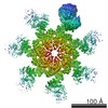
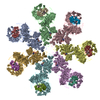
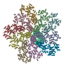
ジャーナル: Genes Dev / 年: 2015 タイトル: Atomic structure of the apoptosome: mechanism of cytochrome c- and dATP-mediated activation of Apaf-1. 著者: Mengying Zhou / Yini Li / Qi Hu / Xiao-Chen Bai / Weiyun Huang / Chuangye Yan / Sjors H W Scheres / Yigong Shi / 要旨: The apoptotic protease-activating factor 1 (Apaf-1) controls the onset of many known forms of intrinsic apoptosis in mammals. Apaf-1 exists in normal cells as an autoinhibited monomer. Upon binding ...The apoptotic protease-activating factor 1 (Apaf-1) controls the onset of many known forms of intrinsic apoptosis in mammals. Apaf-1 exists in normal cells as an autoinhibited monomer. Upon binding to cytochrome c and dATP, Apaf-1 oligomerizes into a heptameric complex known as the apoptosome, which recruits and activates cell-killing caspases. Here we present an atomic structure of an intact mammalian apoptosome at 3.8 Å resolution, determined by single-particle, cryo-electron microscopy (cryo-EM). Structural analysis, together with structure-guided biochemical characterization, uncovered how cytochrome c releases the autoinhibition of Apaf-1 through specific interactions with the WD40 repeats. Structural comparison with autoinhibited Apaf-1 revealed how dATP binding triggers a set of conformational changes that results in the formation of the apoptosome. Together, these results constitute the molecular mechanism of cytochrome c- and dATP-mediated activation of Apaf-1.
 ムービー
ムービー コントローラー


 データを開く
 基本情報
基本情報 要素
要素 キーワード
キーワード 機能・相同性情報
 Homo sapiens (ヒト)
 データ登録者
 引用
引用

 構造の表示
構造の表示 ダウンロードとリンク
 その他のダウンロード

 PDBj
PDBj


 集合体
集合体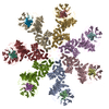
 Trichoplusia ni (イラクサキンウワバ) / 参照: UniProt: O14727
 分子量: 491.182 Da / 分子数: 7 / 由来タイプ: 合成 / 式: C10H16N5O12P3
 分子量: 24.305 Da / 分子数: 7 / 由来タイプ: 合成 / 式: Mg
 分子量: 616.487 Da / 分子数: 7 / 由来タイプ: 合成 / 式: C34H32FeN4O4
 試料調製
試料調製 電子顕微鏡撮影
 FIELD EMISSION GUN / 加速電圧: 300 kV / 照射モード: SPOT SCAN
 解析
解析

