 Movie
Movie Controller


+ Open data

- Basic information
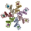
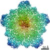
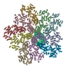
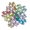
| Entry | Database: PDB / ID: 3j2t | |||||||||
|---|---|---|---|---|---|---|---|---|---|---|
| Title | An improved model of the human apoptosome | |||||||||
 Components Components |
| |||||||||
 Keywords Keywords | APOPTOSIS / Apoptosis protease activating factor-1 / Apaf-1 / cytochrome c | |||||||||
| Function / homology |  Function and homology information Function and homology informationRelease of apoptotic factors from the mitochondria / Formation of apoptosome / Activation of caspases through apoptosome-mediated cleavage / Pyroptosis / Regulation of the apoptosome activity / Transcriptional activation of mitochondrial biogenesis / response to G1 DNA damage checkpoint signaling / Formation of apoptosome / Detoxification of Reactive Oxygen Species / apoptosome ...Release of apoptotic factors from the mitochondria / Formation of apoptosome / Activation of caspases through apoptosome-mediated cleavage / Pyroptosis / Regulation of the apoptosome activity / Transcriptional activation of mitochondrial biogenesis / response to G1 DNA damage checkpoint signaling / Formation of apoptosome / Detoxification of Reactive Oxygen Species / apoptosome / TP53 Regulates Metabolic Genes / Cytoprotection by HMOX1 / cysteine-type endopeptidase activator activity / Respiratory electron transport / Activation of caspases through apoptosome-mediated cleavage / SMAC (DIABLO) binds to IAPs / SMAC(DIABLO)-mediated dissociation of IAP:caspase complexes / Regulation of the apoptosome activity / mitochondrial electron transport, cytochrome c to oxygen / mitochondrial electron transport, ubiquinol to cytochrome c / cysteine-type endopeptidase activator activity involved in apoptotic process / TP53 Regulates Transcription of Caspase Activators and Caspases / Transcriptional Regulation by E2F6 / intrinsic apoptotic signaling pathway in response to endoplasmic reticulum stress / cellular response to transforming growth factor beta stimulus / cardiac muscle cell apoptotic process / heat shock protein binding / response to nutrient / intrinsic apoptotic signaling pathway / kidney development / ADP binding / mitochondrial intermembrane space / nervous system development / secretory granule lumen / regulation of apoptotic process / ficolin-1-rich granule lumen / response to hypoxia / cell differentiation / electron transfer activity / positive regulation of apoptotic process / nucleotide binding / heme binding / apoptotic process / Neutrophil degranulation / protein-containing complex / extracellular exosome / extracellular region / ATP binding / metal ion binding / identical protein binding / nucleus / cytosol Similarity search - Function | |||||||||
| Biological species |  Homo sapiens (human) Homo sapiens (human) | |||||||||
| Method | ELECTRON MICROSCOPY / single particle reconstruction / cryo EM / Resolution: 9.5 Å | |||||||||
 Authors Authors | Yuan, S. / Topf, M. / Akey, C.W. | |||||||||
 Citation Citation | Journal: Biochemistry / Year: 2013 Title: Changes in Apaf-1 conformation that drive apoptosome assembly. Authors: Shujun Yuan / Maya Topf / Thomas F Reubold / Susanne Eschenburg / Christopher W Akey /  Abstract: Apoptosome assembly is highly regulated in the intrinsic cell death pathway. To better understand this step, we created an improved model of the human apoptosome using a crystal structure of full ...Apoptosome assembly is highly regulated in the intrinsic cell death pathway. To better understand this step, we created an improved model of the human apoptosome using a crystal structure of full length Apaf-1 and a single particle, electron density map at ~9.5 Å resolution. The apoptosome model includes N-terminal domains of Apaf-1, cognate β-propellers, and cytochrome c. A direct comparison of Apaf-1 in the apoptosome and as a monomer reveals conformational changes that occur during the first two steps of assembly. This includes an induced-fit mechanism for cytochrome c binding to regulatory β-propellers, which is dependent on shape and charge complementarity, and a large rotation of the nucleotide binding module during nucleotide exchange. These linked conformational changes create an extended Apaf-1 monomer and drive apoptosome assembly. Moreover, the N-terminal CARD in the inactive Apaf-1 monomer is not shielded from other proteins by β-propellers. Hence, the Apaf-1 CARD may be free to interact with a procaspase-9 CARD either before or during apoptosome assembly. Irrespective of the timing, the end product of assembly is a holo-apoptosome with an acentric CARD-CARD disk and tethered pc-9 catalytic domains. Subsequent activation of pc-9 leads to a proteolytic cascade and cell death. #1: Journal: Structure / Year: 2010Title: Structure of an apoptosome-procaspase-9 CARD complex. Authors: Shujun Yuan / Xinchao Yu / Maya Topf / Steven J Ludtke / Xiaodong Wang / Christopher W Akey / Abstract: Apaf-1 coassembles with cytochrome c to form the apoptosome, which then binds and activates procaspase-9 (pc-9). We removed pc-9 catalytic domains from the holoapoptosome by site-directed ...Apaf-1 coassembles with cytochrome c to form the apoptosome, which then binds and activates procaspase-9 (pc-9). We removed pc-9 catalytic domains from the holoapoptosome by site-directed thrombinolysis. A structure of the resulting apoptosome-pc-9 CARD complex was then determined at approximately 9.5 A resolution. In our model, the central hub is constructed like other AAA+ protein rings but also contains novel features. At higher radius, the regulatory region of each Apaf-1 is comprised of tandem seven and eight blade beta-propellers with cytochrome c docked between them. Remarkably, Apaf-1 CARDs are disordered in the ground state. During activation, each Apaf-1 CARD interacts with a pc-9 CARD and these heterodimers form a flexibly tethered "disk" that sits above the central hub. When taken together, the data reveal conformational changes during Apaf-1 assembly that allow pc-9 activation. The model also provides a plausible explanation for the effects of NOD mutations that have been mapped onto the central hub. | |||||||||
| History |
|
- Structure visualization
| Movie |
Movie viewer |
|---|---|
| Structure viewer | Molecule: MolmilJmol/JSmol |
- Downloads & links
-Download
| PDBx/mmCIF format | 3j2t.cif.gz | 1.5 MB | Display | PDBx/mmCIF format |
|---|---|---|---|---|
| PDB format | pdb3j2t.ent.gz | 1.2 MB | Display | PDB format |
| PDBx/mmJSON format | 3j2t.json.gz | Tree view | PDBx/mmJSON format | |
| Others |  Other downloads Other downloads |
-Validation report
| Arichive directory | https://data.pdbj.org/pub/pdb/validation_reports/j2/3j2tftp://data.pdbj.org/pub/pdb/validation_reports/j2/3j2t | HTTPS FTP |
|---|
-Related structure data
| Related structure data | 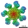 5186M M: map data used to model this data |
|---|---|
| Similar structure data |

-Links
 PDBj
PDBj


- Assembly
| Deposited unit | 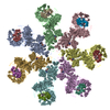
|
|---|---|
| 1 |
|
-Components
| #1: Protein | Mass: 144095.812 Da / Num. of mol.: 7 Source method: isolated from a genetically manipulated source Source: (gene. exp.) Homo sapiens (human) / Gene: Apaf-1, APAF1, KIAA0413 / Plasmid: pFastBac1 / Production host:   Spodoptera frugiperda (fall armyworm) / Strain (production host): sf21 / References: UniProt: O14727 Spodoptera frugiperda (fall armyworm) / Strain (production host): sf21 / References: UniProt: O14727#2: Protein | Mass: 11595.392 Da / Num. of mol.: 7 / Source method: isolated from a natural source / Source: (natural) #3: Chemical | ChemComp-ATP /   Mass: 507.181 Da / Num. of mol.: 7 / Source method: obtained synthetically / Formula: C10H16N5O13P3 / Comment: ATP, energy-carrying molecule*YM Mass: 507.181 Da / Num. of mol.: 7 / Source method: obtained synthetically / Formula: C10H16N5O13P3 / Comment: ATP, energy-carrying molecule*YM#4: Chemical | ChemComp-HEM /   Mass: 616.487 Da / Num. of mol.: 7 / Source method: obtained synthetically / Formula: C34H32FeN4O4 Mass: 616.487 Da / Num. of mol.: 7 / Source method: obtained synthetically / Formula: C34H32FeN4O4Has protein modification | Y | |
|---|
-Experimental details
-Experiment
| Experiment | Method: ELECTRON MICROSCOPY |
|---|---|
| EM experiment | Aggregation state: PARTICLE / 3D reconstruction method: single particle reconstruction |
- Sample preparation
| Component |
| |||||||||||||||
|---|---|---|---|---|---|---|---|---|---|---|---|---|---|---|---|---|
| Buffer solution | Name: low salt HEPES buffer / pH: 7.5 Details: 20mM HEPES, 10mM KCl, 1.5mM MgCl2, 1mM EDTA, 1mM EGTA, 1mM DTT | |||||||||||||||
| Specimen | Conc.: 3 mg/ml / Embedding applied: NO / Shadowing applied: NO / Staining applied: NO / Vitrification applied: YES | |||||||||||||||
| Specimen support | Details: Quantifoil R1.2/1.3 holey grid | |||||||||||||||
| Vitrification | Instrument: FEI VITROBOT MARK III / Cryogen name: ETHANE / Temp: 77 K / Humidity: 100 % Details: Blot for 2 seconds before plunging (FEI VITROBOT MARK III) Method: Blot for 2 seconds before plunging |
- Electron microscopy imaging
| Experimental equipment |  Model: Tecnai F20 / Image courtesy: FEI Company |
|---|---|
| Microscopy | Model: FEI TECNAI F20 / Date: Aug 1, 2008 |
| Electron gun | Electron source:  FIELD EMISSION GUN / Accelerating voltage: 120 kV / Illumination mode: FLOOD BEAM FIELD EMISSION GUN / Accelerating voltage: 120 kV / Illumination mode: FLOOD BEAM |
| Electron lens | Mode: BRIGHT FIELD / Nominal magnification: 62000 X / Calibrated magnification: 62000 X / Nominal defocus max: 3000 nm / Nominal defocus min: 1500 nm / Cs: 2 mm |
| Specimen holder | Specimen holder model: GATAN LIQUID NITROGEN Specimen holder type: Side entry liquid nitrogen-cooled cryo specimen holder Temperature: 96 K / Tilt angle max: 0 ° / Tilt angle min: 0 ° |
| Image recording | Electron dose: 25 e/Å2 / Film or detector model: KODAK SO-163 FILM |
| Image scans | Num. digital images: 400 |
| Radiation | Protocol: SINGLE WAVELENGTH / Monochromatic (M) / Laue (L): M / Scattering type: x-ray |
| Radiation wavelength | Relative weight: 1 |
- Processing
| EM software |
| ||||||||||||||||||||||||||||
|---|---|---|---|---|---|---|---|---|---|---|---|---|---|---|---|---|---|---|---|---|---|---|---|---|---|---|---|---|---|
| CTF correction | Details: CTF correction was done on each particle image based on summed power spectra from each micrograph. | ||||||||||||||||||||||||||||
| Symmetry | Point symmetry: C7 (7 fold cyclic) | ||||||||||||||||||||||||||||
| 3D reconstruction | Method: Projection Matching / Resolution: 9.5 Å / Num. of particles: 34000 / Nominal pixel size: 2.26 Å / Actual pixel size: 2.26 Å / Magnification calibration: TMV images Details: EMAN was used for reconstruction using C7 symmetry. Symmetry type: POINT | ||||||||||||||||||||||||||||
| Atomic model building |
| ||||||||||||||||||||||||||||
| Atomic model building |
| ||||||||||||||||||||||||||||
| Refinement step | Cycle: LAST
|


