response to G1 DNA damage checkpoint signaling / apoptosome / Formation of apoptosome / cytochrome complex / cysteine-type endopeptidase activator activity / Activation of caspases through apoptosome-mediated cleavage / SMAC (DIABLO) binds to IAPs / SMAC(DIABLO)-mediated dissociation of IAP:caspase complexes / Regulation of the apoptosome activity / mitochondrial electron transport, cytochrome c to oxygen ...response to G1 DNA damage checkpoint signaling / apoptosome / Formation of apoptosome / cytochrome complex / cysteine-type endopeptidase activator activity / Activation of caspases through apoptosome-mediated cleavage / SMAC (DIABLO) binds to IAPs / SMAC(DIABLO)-mediated dissociation of IAP:caspase complexes / Regulation of the apoptosome activity / mitochondrial electron transport, cytochrome c to oxygen / mitochondrial electron transport, ubiquinol to cytochrome c / cysteine-type endopeptidase activator activity involved in apoptotic process / TP53 Regulates Transcription of Caspase Activators and Caspases / Transcriptional Regulation by E2F6 / intrinsic apoptotic signaling pathway in response to endoplasmic reticulum stress / cellular response to transforming growth factor beta stimulus / cardiac muscle cell apoptotic process / heat shock protein binding / kidney development / response to nutrient / intrinsic apoptotic signaling pathway / apoptotic signaling pathway / ADP binding / mitochondrial intermembrane space / nervous system development / secretory granule lumen / regulation of apoptotic process / ficolin-1-rich granule lumen / response to hypoxia / electron transfer activity / cell differentiation / positive regulation of apoptotic process / nucleotide binding / heme binding / apoptotic process / Neutrophil degranulation / lipid binding / protein-containing complex / extracellular exosome / extracellular region / ATP binding / metal ion binding / identical protein binding / nucleus / cytosol Similarity search - Function
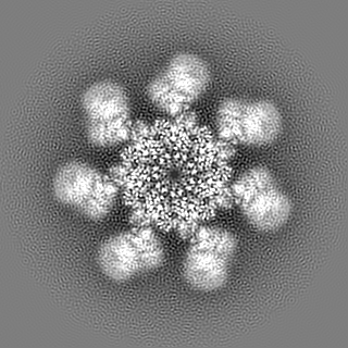
Journal: Genes Dev / Year: 2015 Title: Atomic structure of the apoptosome: mechanism of cytochrome c- and dATP-mediated activation of Apaf-1. Authors: Mengying Zhou / Yini Li / Qi Hu / Xiao-Chen Bai / Weiyun Huang / Chuangye Yan / Sjors H W Scheres / Yigong Shi / Abstract: The apoptotic protease-activating factor 1 (Apaf-1) controls the onset of many known forms of intrinsic apoptosis in mammals. Apaf-1 exists in normal cells as an autoinhibited monomer. Upon binding ...The apoptotic protease-activating factor 1 (Apaf-1) controls the onset of many known forms of intrinsic apoptosis in mammals. Apaf-1 exists in normal cells as an autoinhibited monomer. Upon binding to cytochrome c and dATP, Apaf-1 oligomerizes into a heptameric complex known as the apoptosome, which recruits and activates cell-killing caspases. Here we present an atomic structure of an intact mammalian apoptosome at 3.8 Å resolution, determined by single-particle, cryo-electron microscopy (cryo-EM). Structural analysis, together with structure-guided biochemical characterization, uncovered how cytochrome c releases the autoinhibition of Apaf-1 through specific interactions with the WD40 repeats. Structural comparison with autoinhibited Apaf-1 revealed how dATP binding triggers a set of conformational changes that results in the formation of the apoptosome. Together, these results constitute the molecular mechanism of cytochrome c- and dATP-mediated activation of Apaf-1.
History
Deposition
Oct 14, 2015
-
Header (metadata) release
Nov 18, 2015
-
Map release
Nov 18, 2015
-
Update
Nov 18, 2015
-
Current status
Nov 18, 2015
Processing site: PDBj / Status: Released
-
Structure visualization
Movie
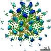
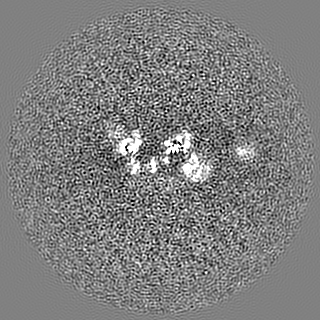
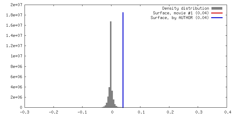
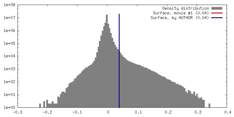
Surface view with section colored by density value
Entire : Heptameric apoptosome of activated human Apaf-1 binding to horse ...
Entire
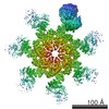
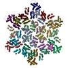
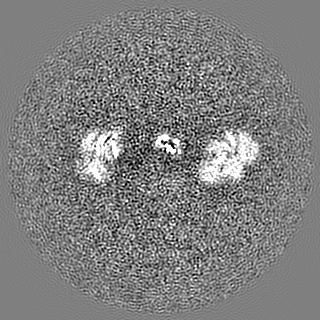
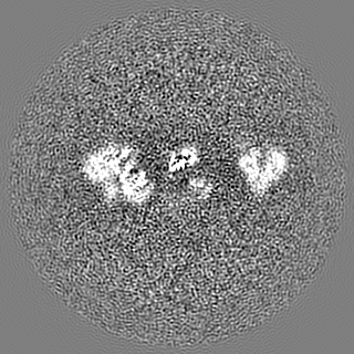
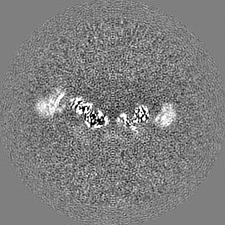
Name: Heptameric apoptosome of activated human Apaf-1 binding to horse cytochrome c and dATP
Components
Sample: Heptameric apoptosome of activated human Apaf-1 binding to horse cytochrome c and dATP
Protein or peptide: The apoptotic protease activating factor 1
Protein or peptide: cytochrome c
-
Supramolecule #1000: Heptameric apoptosome of activated human Apaf-1 binding to horse ...
Supramolecule
Name: Heptameric apoptosome of activated human Apaf-1 binding to horse cytochrome c and dATP type: sample / ID: 1000 / Oligomeric state: Heptamer / Number unique components: 2
-
Macromolecule #1: The apoptotic protease activating factor 1
Macromolecule
Name: The apoptotic protease activating factor 1 / type: protein_or_peptide / ID: 1 / Name.synonym: Apaf-1 / Number of copies: 7 / Oligomeric state: Heptamer / Recombinant expression: Yes
Source (natural)
Organism: Homo sapiens (human) / synonym: Human
Recombinant expression
Organism: Trichoplusia ni (cabbage looper) / Recombinant cell: High Five / Recombinant plasmid: pFastBac
Sequence
UniProtKB: Apoptotic protease-activating factor 1
-
Macromolecule #2: cytochrome c
Macromolecule
Name: cytochrome c / type: protein_or_peptide / ID: 2 / Number of copies: 7 / Recombinant expression: No / Database: NCBI
Source (natural)
Organism: Equus caballus (horse) / synonym: Horse
Sequence
UniProtKB: Cytochrome c
-
Experimental details
-
Structure determination
Method
cryo EM
Processing
single particle reconstruction
Aggregation state
particle
-
Sample preparation
Buffer
pH: 7.5 Details: 20 mM HEPES, 10 mM KCl, 1.5 mM MgCl2, 1 mM EDTA and 1 mM DTT
Grid
Details: 400-mesh Cu R 1.2/1.3 grids
Vitrification
Cryogen name: ETHANE / Chamber humidity: 100 % / Instrument: FEI VITROBOT MARK IV / Method: Blot for 2.5 seconds before plunging
-
Electron microscopy
Microscope
FEI TITAN KRIOS
Date
Jan 10, 2015
Image recording
Category: CCD / Film or detector model: GATAN K2 (4k x 4k) / Number real images: 912 / Average electron dose: 40 e/Å2
Electron beam
Acceleration voltage: 300 kV / Electron source: FIELD EMISSION GUN
In the structure databanks used in Yorodumi, some data are registered as the other names, "COVID-19 virus" and "2019-nCoV". Here are the details of the virus and the list of structure data.
Jan 31, 2019. EMDB accession codes are about to change! (news from PDBe EMDB page)
EMDB accession codes are about to change! (news from PDBe EMDB page)
The allocation of 4 digits for EMDB accession codes will soon come to an end. Whilst these codes will remain in use, new EMDB accession codes will include an additional digit and will expand incrementally as the available range of codes is exhausted. The current 4-digit format prefixed with “EMD-” (i.e. EMD-XXXX) will advance to a 5-digit format (i.e. EMD-XXXXX), and so on. It is currently estimated that the 4-digit codes will be depleted around Spring 2019, at which point the 5-digit format will come into force.
The EM Navigator/Yorodumi systems omit the EMD- prefix.
Related info.:Q: What is EMD? / ID/Accession-code notation in Yorodumi/EM Navigator
Yorodumi is a browser for structure data from EMDB, PDB, SASBDB, etc.
This page is also the successor to EM Navigator detail page, and also detail information page/front-end page for Omokage search.
The word "yorodu" (or yorozu) is an old Japanese word meaning "ten thousand". "mi" (miru) is to see.
Related info.:EMDB / PDB / SASBDB / Comparison of 3 databanks / Yorodumi Search / Aug 31, 2016. New EM Navigator & Yorodumi / Yorodumi Papers / Jmol/JSmol / Function and homology information / Changes in new EM Navigator and Yorodumi
 Movie
Movie Controller


 Open data
 Basic information
 Map data
 Sample
 Keywords
 Function and homology information
 Homo sapiens (human) /
 Authors
 Citation

 Structure visualization
 Downloads & links
 400_6480.gif
 80_6480.gif
 http://ftp.pdbj.org/pub/emdb/structures/EMD-6480


 Z (Sec.)
 Y (Row.)
 X (Col.)


 Sample components
 Trichoplusia ni (cabbage looper) / Recombinant cell: High Five / Recombinant plasmid: pFastBac
 Processing
 Electron microscopy
 FIELD EMISSION GUN

