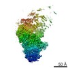
Movie
Movie Controller

[English] 日本語
 Yorodumi
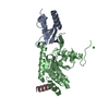
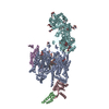
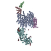
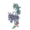
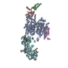
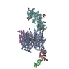
- PDB-3jbr: Cryo-EM structure of the rabbit voltage-gated calcium channel Cav... -
+ Open data

- Basic information
| Entry | Database: PDB / ID: 3jbr | ||||||
|---|---|---|---|---|---|---|---|
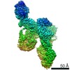
| Title | Cryo-EM structure of the rabbit voltage-gated calcium channel Cav1.1 complex at 4.2 angstrom | ||||||
 Components Components |
| ||||||
 Keywords Keywords | MEMBRANE PROTEIN / voltage-gated calcium channel | ||||||
| Function / homology |  Function and homology information Function and homology informationvoltage-gated calcium channel activity involved in regulation of presynaptic cytosolic calcium levels / Phase 0 - rapid depolarisation / Phase 2 - plateau phase / positive regulation of calcium ion transmembrane transport via high voltage-gated calcium channel / Regulation of insulin secretion / Presynaptic depolarization and calcium channel opening / membrane depolarization during atrial cardiac muscle cell action potential / high voltage-gated calcium channel activity / photoreceptor ribbon synapse / membrane depolarization during AV node cell action potential ...voltage-gated calcium channel activity involved in regulation of presynaptic cytosolic calcium levels / Phase 0 - rapid depolarisation / Phase 2 - plateau phase / positive regulation of calcium ion transmembrane transport via high voltage-gated calcium channel / Regulation of insulin secretion / Presynaptic depolarization and calcium channel opening / membrane depolarization during atrial cardiac muscle cell action potential / high voltage-gated calcium channel activity / photoreceptor ribbon synapse / membrane depolarization during AV node cell action potential / L-type voltage-gated calcium channel complex / positive regulation of muscle contraction / positive regulation of calcium ion transport / calcium ion import / regulation of calcium ion transmembrane transport via high voltage-gated calcium channel / voltage-gated calcium channel complex / cellular response to caffeine / regulation of heart rate by cardiac conduction / calcium ion import across plasma membrane / regulation of ryanodine-sensitive calcium-release channel activity / voltage-gated calcium channel activity / release of sequestered calcium ion into cytosol / visual perception / T-tubule / muscle contraction / protein localization to plasma membrane / calcium channel regulator activity / phosphoprotein binding / sarcolemma / calcium ion transmembrane transport / actin filament binding / calcium ion transport / presynapse / chemical synaptic transmission / transmembrane transporter binding / calmodulin binding / protein domain specific binding / protein kinase binding / metal ion binding / identical protein binding / plasma membrane Similarity search - Function | ||||||
| Biological species |  | ||||||
| Method | ELECTRON MICROSCOPY / single particle reconstruction / cryo EM / Resolution: 4.2 Å | ||||||
 Authors Authors | Wu, J.P. / Yan, Z. / Yan, N. | ||||||
 Citation Citation | Journal: Science / Year: 2015 Title: Structure of the voltage-gated calcium channel Cav1.1 complex. Authors: Jianping Wu / Zhen Yan / Zhangqiang Li / Chuangye Yan / Shan Lu / Mengqiu Dong / Nieng Yan /  Abstract: The voltage-gated calcium channel Ca(v)1.1 is engaged in the excitation-contraction coupling of skeletal muscles. The Ca(v)1.1 complex consists of the pore-forming subunit α1 and auxiliary subunits ...The voltage-gated calcium channel Ca(v)1.1 is engaged in the excitation-contraction coupling of skeletal muscles. The Ca(v)1.1 complex consists of the pore-forming subunit α1 and auxiliary subunits α2δ, β, and γ. We report the structure of the rabbit Ca(v)1.1 complex determined by single-particle cryo-electron microscopy. The four homologous repeats of the α1 subunit are arranged clockwise in the extracellular view. The γ subunit, whose structure resembles claudins, interacts with the voltage-sensing domain of repeat IV (VSD(IV)), whereas the cytosolic β subunit is located adjacent to VSD(II) of α1. The α2 subunit interacts with the extracellular loops of repeats I to III through its VWA and Cache1 domains. The structure reveals the architecture of a prototypical eukaryotic Ca(v) channel and provides a framework for understanding the function and disease mechanisms of Ca(v) and Na(v) channels. | ||||||
| History |
|
- Structure visualization
| Movie |
Movie viewer |
|---|---|
| Structure viewer | Molecule: MolmilJmol/JSmol |
- Downloads & links
-Download
| PDBx/mmCIF format | 3jbr.cif.gz | 454 KB | Display | PDBx/mmCIF format |
|---|---|---|---|---|
| PDB format | pdb3jbr.ent.gz | 335.5 KB | Display | PDB format |
| PDBx/mmJSON format | 3jbr.json.gz | Tree view | PDBx/mmJSON format | |
| Others |  Other downloads Other downloads |
-Validation report
| Summary document | 3jbr_validation.pdf.gz | 982.3 KB | Display | wwPDB validaton report |
|---|---|---|---|---|
| Full document | 3jbr_full_validation.pdf.gz | 1 MB | Display | |
| Data in XML | 3jbr_validation.xml.gz | 65.6 KB | Display | |
| Data in CIF | 3jbr_validation.cif.gz | 103.5 KB | Display | |
| Arichive directory | https://data.pdbj.org/pub/pdb/validation_reports/jb/3jbrftp://data.pdbj.org/pub/pdb/validation_reports/jb/3jbr | HTTPS FTP |
-Related structure data
| Related structure data | 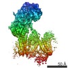 6475MC 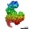 6476MC M: map data used to model this data C: citing same article ( |
|---|---|
| Similar structure data |


-Links
 PDBj
PDBj


- Assembly
| Deposited unit | 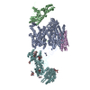
|
|---|---|
| 1 |
|
-Components
-Voltage-dependent L-type calcium channel subunit ... , 2 types, 2 molecules AB
| #1: Protein | Mass: 208655.859 Da / Num. of mol.: 1 / Source method: isolated from a natural source / Source: (natural) |
|---|---|
| #2: Protein | Mass: 40300.004 Da / Num. of mol.: 1 Source method: isolated from a genetically manipulated source Details: Chain B is for a subunit that is docked by a homolog crystal structure (PDB code: 1T0J) Source: (gene. exp.)  |
-Voltage-dependent calcium channel ... , 2 types, 2 molecules EF
| #3: Protein | Mass: 25082.254 Da / Num. of mol.: 1 / Source method: isolated from a natural source / Source: (natural) |
|---|---|
| #4: Protein | Mass: 116775.609 Da / Num. of mol.: 1 / Source method: isolated from a natural source / Source: (natural) |
-Sugars , 2 types, 17 molecules 

| #6: Sugar | ChemComp-NAG /  Type: D-saccharide, beta linking / Mass: 221.208 Da / Num. of mol.: 15 Type: D-saccharide, beta linking / Mass: 221.208 Da / Num. of mol.: 15Source method: isolated from a genetically manipulated source Formula: C8H15NO6 #7: Sugar |  Type: D-saccharide, beta linking / Mass: 180.156 Da / Num. of mol.: 2 Type: D-saccharide, beta linking / Mass: 180.156 Da / Num. of mol.: 2Source method: isolated from a genetically manipulated source Formula: C6H12O6 |
|---|
-Non-polymers , 1 types, 1 molecules 
| #5: Chemical | ChemComp-CA /  Mass: 40.078 Da / Num. of mol.: 1 / Source method: obtained synthetically / Formula: Ca Mass: 40.078 Da / Num. of mol.: 1 / Source method: obtained synthetically / Formula: Ca |
|---|
-Details
| Has protein modification | Y |
|---|---|
| Sequence details | RESIDUES 1396-1520 OF ENTITY 1 (CHAIN A), AND RESIDUES 40-55/662-955 OF ENTITY 4 (CHAIN F) ARE ...RESIDUES 1396-1520 OF ENTITY 1 (CHAIN A), AND RESIDUES 40-55/662-955 OF ENTITY 4 (CHAIN F) ARE MODELED AS UNK SINCE THE EM DENSITY IS NOT GOOD ENOUGH TO ASSIGN EXACTLY. THE REAL SEQUENCE OF RESIDUES 1396-1520 OF ENTITY 1 (CHAIN A) SHOULD BE (PHHLDEFKAI |
-Experimental details
-Experiment
| Experiment | Method: ELECTRON MICROSCOPY |
|---|---|
| EM experiment | Aggregation state: PARTICLE / 3D reconstruction method: single particle reconstruction |
- Sample preparation
| Component | Name: a membrane protein cryo-sample / Type: COMPLEX / Synonym: Cav1.1 |
|---|---|
| Molecular weight | Value: 0.45 MDa / Experimental value: NO |
| Buffer solution | Name: 20mM MOPS, 200mM NaCl, 0.5mM CaCl2,0.1% digitonin / pH: 7.4 / Details: 20mM MOPS, 200mM NaCl, 0.5mM CaCl2,0.1% digitonin |
| Specimen | Conc.: 0.1 mg/ml / Embedding applied: NO / Shadowing applied: NO / Staining applied: NO / Vitrification applied: YES |
| Specimen support | Details: carbon coated grid |
| Vitrification | Instrument: FEI VITROBOT MARK IV / Cryogen name: ETHANE / Humidity: 100 % / Method: Blot for 2 seconds before plunging |
- Electron microscopy imaging
| Experimental equipment |  Model: Titan Krios / Image courtesy: FEI Company |
|---|---|
| Microscopy | Model: FEI TITAN KRIOS / Date: Feb 2, 2015 |
| Electron gun | Electron source:  FIELD EMISSION GUN / Accelerating voltage: 300 kV / Illumination mode: SPOT SCAN FIELD EMISSION GUN / Accelerating voltage: 300 kV / Illumination mode: SPOT SCAN |
| Electron lens | Mode: BRIGHT FIELD / Nominal defocus max: 3.3 nm / Nominal defocus min: 2 nm / Cs: 2.7 mm / Camera length: 0 mm |
| Specimen holder | Specimen holder model: FEI TITAN KRIOS AUTOGRID HOLDER / Tilt angle max: 0 ° / Tilt angle min: 0 ° |
| Image recording | Electron dose: 50 e/Å2 / Film or detector model: GATAN K2 (4k x 4k) |
- Processing
| EM software |
| ||||||||||||
|---|---|---|---|---|---|---|---|---|---|---|---|---|---|
| CTF correction | Details: each micrograph | ||||||||||||
| Symmetry | Point symmetry: C1 (asymmetric) | ||||||||||||
| 3D reconstruction | Resolution: 4.2 Å / Resolution method: FSC 0.143 CUT-OFF / Num. of particles: 353372 Details: (Single particle details: The particles were selected using RELION.) (Single particle--Applied symmetry: C1) Symmetry type: POINT | ||||||||||||
| Atomic model building | Protocol: RIGID BODY FIT / Space: REAL Details: REFINEMENT PROTOCOL--rigid body DETAILS--The fitting is mostly refer to a related entry | ||||||||||||
| Atomic model building | PDB-ID: 1T0J Accession code: 1T0J / Source name: PDB / Type: experimental model | ||||||||||||
| Refinement step | Cycle: LAST
|