 Movie
Movie Controller

[English] 日本語
 Yorodumi
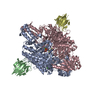
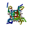
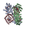
- PDB-6byo: Residue assignment correction to the voltage gated calcium Cav1.1... -
+ Open data

- Basic information
| Entry | Database: PDB / ID: 6byo | |||||||||
|---|---|---|---|---|---|---|---|---|---|---|
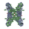
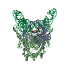
| Title | Residue assignment correction to the voltage gated calcium Cav1.1 rabbit alpha 1 subunit PDB entries 3JBR & 5GJV | |||||||||
 Components Components | Voltage-dependent L-type calcium channel subunit alpha-1S | |||||||||
 Keywords Keywords | MEMBRANE PROTEIN / Voltage-Gated Calcium Channels | |||||||||
| Function / homology |  Function and homology information Function and homology informationhigh voltage-gated calcium channel activity / L-type voltage-gated calcium channel complex / positive regulation of muscle contraction / regulation of release of sequestered calcium ion into cytosol / cellular response to caffeine / calcium ion import across plasma membrane / voltage-gated calcium channel activity / release of sequestered calcium ion into cytosol / muscle contraction / T-tubule ...high voltage-gated calcium channel activity / L-type voltage-gated calcium channel complex / positive regulation of muscle contraction / regulation of release of sequestered calcium ion into cytosol / cellular response to caffeine / calcium ion import across plasma membrane / voltage-gated calcium channel activity / release of sequestered calcium ion into cytosol / muscle contraction / T-tubule / calcium ion transmembrane transport / transmembrane transporter binding / calmodulin binding / metal ion binding / plasma membrane Similarity search - Function | |||||||||
| Biological species |  | |||||||||
| Method | ELECTRON MICROSCOPY / single particle reconstruction / cryo EM / Resolution: 3.6 Å | |||||||||
 Authors Authors | Cardozo, T.J. / Martinez-Ortiz, W. | |||||||||
| Funding support |  United States, 2items United States, 2items
| |||||||||
 Citation Citation | Journal: Nature / Year: 2016 Title: Structure of the voltage-gated calcium channel Ca(v)1.1 at 3.6 Å resolution. Authors: Jianping Wu / Zhen Yan / Zhangqiang Li / Xingyang Qian / Shan Lu / Mengqiu Dong / Qiang Zhou / Nieng Yan /  Abstract: The voltage-gated calcium (Ca) channels convert membrane electrical signals to intracellular Ca-mediated events. Among the ten subtypes of Ca channel in mammals, Ca1.1 is specified for the excitation- ...The voltage-gated calcium (Ca) channels convert membrane electrical signals to intracellular Ca-mediated events. Among the ten subtypes of Ca channel in mammals, Ca1.1 is specified for the excitation-contraction coupling of skeletal muscles. Here we present the cryo-electron microscopy structure of the rabbit Ca1.1 complex at a nominal resolution of 3.6 Å. The inner gate of the ion-conducting α1-subunit is closed and all four voltage-sensing domains adopt an 'up' conformation, suggesting a potentially inactivated state. The extended extracellular loops of the pore domain, which are stabilized by multiple disulfide bonds, form a windowed dome above the selectivity filter. One side of the dome provides the docking site for the α2δ-1-subunit, while the other side may attract cations through its negative surface potential. The intracellular I-II and III-IV linker helices interact with the β-subunit and the carboxy-terminal domain of α1, respectively. Classification of the particles yielded two additional reconstructions that reveal pronounced displacement of β and adjacent elements in α1. The atomic model of the Ca1.1 complex establishes a foundation for mechanistic understanding of excitation-contraction coupling and provides a three-dimensional template for molecular interpretations of the functions and disease mechanisms of Ca and Na channels. #1: Journal: Science / Year: 2015Title: Structure of the voltage-gated calcium channel Cav1.1 complex. Authors: Jianping Wu / Zhen Yan / Zhangqiang Li / Chuangye Yan / Shan Lu / Mengqiu Dong / Nieng Yan / Abstract: The voltage-gated calcium channel Ca(v)1.1 is engaged in the excitation-contraction coupling of skeletal muscles. The Ca(v)1.1 complex consists of the pore-forming subunit α1 and auxiliary subunits ...The voltage-gated calcium channel Ca(v)1.1 is engaged in the excitation-contraction coupling of skeletal muscles. The Ca(v)1.1 complex consists of the pore-forming subunit α1 and auxiliary subunits α2δ, β, and γ. We report the structure of the rabbit Ca(v)1.1 complex determined by single-particle cryo-electron microscopy. The four homologous repeats of the α1 subunit are arranged clockwise in the extracellular view. The γ subunit, whose structure resembles claudins, interacts with the voltage-sensing domain of repeat IV (VSD(IV)), whereas the cytosolic β subunit is located adjacent to VSD(II) of α1. The α2 subunit interacts with the extracellular loops of repeats I to III through its VWA and Cache1 domains. The structure reveals the architecture of a prototypical eukaryotic Ca(v) channel and provides a framework for understanding the function and disease mechanisms of Ca(v) and Na(v) channels. | |||||||||
| History |
| |||||||||
| Remark 0 | THIS ENTRY 6BYO REFLECTS AN ALTERNATIVE MODELING OF THE ORIGINAL DATA IN EMD-9513, DETERMINED BY J.P.Wu,Z.Yan |
- Structure visualization
| Movie |
Movie viewer |
|---|---|
| Structure viewer | Molecule: MolmilJmol/JSmol |
- Downloads & links
-Download
| PDBx/mmCIF format | 6byo.cif.gz | 438.7 KB | Display | PDBx/mmCIF format |
|---|---|---|---|---|
| PDB format | pdb6byo.ent.gz | 358.1 KB | Display | PDB format |
| PDBx/mmJSON format | 6byo.json.gz | Tree view | PDBx/mmJSON format | |
| Others |  Other downloads Other downloads |
-Validation report
| Arichive directory | https://data.pdbj.org/pub/pdb/validation_reports/by/6byoftp://data.pdbj.org/pub/pdb/validation_reports/by/6byo | HTTPS FTP |
|---|
-Related structure data
| Related structure data | 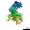 9513M M: map data used to model this data |
|---|---|
| Similar structure data |


-Links
 PDBj
PDBj
- Assembly
| Deposited unit | 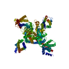
|
|---|---|
| 1 |
|
-Components
| #1: Protein | Mass: 155063.562 Da / Num. of mol.: 1 / Source method: isolated from a natural source / Source: (natural) |
|---|---|
| Has protein modification | Y |
-Experimental details
-Experiment
| Experiment | Method: ELECTRON MICROSCOPY |
|---|---|
| EM experiment | Aggregation state: PARTICLE / 3D reconstruction method: single particle reconstruction |
- Sample preparation
| Component | Name: Rabbit Voltage Gated Calcium Cav1.1 alpha 1 subunit. / Type: COMPLEX / Entity ID: all / Source: RECOMBINANT |
|---|---|
| Source (natural) | Organism: |
| Source (recombinant) | Organism:  |
| Buffer solution | pH: 6.5 |
| Specimen | Embedding applied: NO / Shadowing applied: NO / Staining applied: NO / Vitrification applied: YES |
| Vitrification | Cryogen name: NITROGEN |
- Electron microscopy imaging
| Experimental equipment |  Model: Titan Krios / Image courtesy: FEI Company |
|---|---|
| Microscopy | Model: FEI TITAN KRIOS |
| Electron gun | Electron source:  FIELD EMISSION GUN / Accelerating voltage: 300 kV / Illumination mode: SPOT SCAN FIELD EMISSION GUN / Accelerating voltage: 300 kV / Illumination mode: SPOT SCAN |
| Electron lens | Mode: BRIGHT FIELD |
| Image recording | Electron dose: 50 e/Å2 / Film or detector model: GATAN K2 SUMMIT (4k x 4k) |
- Processing
| EM software | Name: PHENIX / Category: model fitting |
|---|---|
| CTF correction | Type: NONE |
| 3D reconstruction | Resolution: 3.6 Å / Resolution method: OTHER / Num. of particles: 527833 / Details: overall resolution of entire complex / Symmetry type: POINT |
| Atomic model building | Protocol: RIGID BODY FIT |
