 Movie
Movie Controller


+ Open data

- Basic information
| Entry | Database: PDB / ID: 6lqa | ||||||
|---|---|---|---|---|---|---|---|
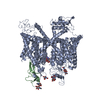
| Title | voltage-gated sodium channel Nav1.5 with quinidine | ||||||
 Components Components | Sodium channel protein type 5 subunit alpha | ||||||
 Keywords Keywords | TRANSPORT PROTEIN / membrane protein / Voltage-gated sodium channel | ||||||
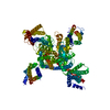
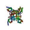
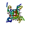
| Function / homology |  Function and homology information Function and homology informationvoltage-gated sodium channel activity involved in AV node cell action potential / voltage-gated sodium channel activity involved in bundle of His cell action potential / voltage-gated sodium channel activity involved in Purkinje myocyte action potential / voltage-gated sodium channel activity involved in SA node cell action potential / bundle of His cell action potential / regulation of ventricular cardiac muscle cell membrane depolarization / AV node cell action potential / SA node cell action potential / AV node cell to bundle of His cell communication / membrane depolarization during SA node cell action potential ...voltage-gated sodium channel activity involved in AV node cell action potential / voltage-gated sodium channel activity involved in bundle of His cell action potential / voltage-gated sodium channel activity involved in Purkinje myocyte action potential / voltage-gated sodium channel activity involved in SA node cell action potential / bundle of His cell action potential / regulation of ventricular cardiac muscle cell membrane depolarization / AV node cell action potential / SA node cell action potential / AV node cell to bundle of His cell communication / membrane depolarization during SA node cell action potential / response to denervation involved in regulation of muscle adaptation / membrane depolarization during atrial cardiac muscle cell action potential / voltage-gated sodium channel activity involved in cardiac muscle cell action potential / regulation of atrial cardiac muscle cell membrane repolarization / cardiac ventricle development / brainstem development / regulation of atrial cardiac muscle cell membrane depolarization / positive regulation of action potential / atrial cardiac muscle cell action potential / membrane depolarization during AV node cell action potential / cardiac conduction system development / membrane depolarization during bundle of His cell action potential / telencephalon development / membrane depolarization during Purkinje myocyte cell action potential / membrane depolarization during cardiac muscle cell action potential / positive regulation of sodium ion transport / membrane depolarization during action potential / regulation of sodium ion transmembrane transport / ventricular cardiac muscle cell action potential / regulation of ventricular cardiac muscle cell membrane repolarization / cardiac muscle cell action potential involved in contraction / voltage-gated sodium channel complex / regulation of cardiac muscle cell contraction / voltage-gated sodium channel activity / Interaction between L1 and Ankyrins / ankyrin binding / odontogenesis of dentin-containing tooth / sodium ion transport / Phase 0 - rapid depolarisation / regulation of heart rate by cardiac conduction / nitric-oxide synthase binding / fibroblast growth factor binding / intercalated disc / lateral plasma membrane / membrane depolarization / cardiac muscle contraction / T-tubule / regulation of heart rate / cellular response to calcium ion / cerebellum development / positive regulation of epithelial cell proliferation / sodium ion transmembrane transport / sarcolemma / caveola / Z disc / scaffold protein binding / transmembrane transporter binding / calmodulin binding / protein domain specific binding / ubiquitin protein ligase binding / protein kinase binding / nucleolus / perinuclear region of cytoplasm / enzyme binding / cell surface / endoplasmic reticulum / nucleoplasm / membrane / plasma membrane Similarity search - Function | ||||||
| Biological species |  Homo sapiens (human) Homo sapiens (human) | ||||||
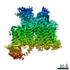
| Method | ELECTRON MICROSCOPY / single particle reconstruction / cryo EM / Resolution: 3.3 Å | ||||||
 Authors Authors | Yan, N. / Li, Z. / Pan, X. / Huang, G. | ||||||
| Funding support |  China, 1items China, 1items
| ||||||
 Citation Citation | Journal: Angew Chem Int Ed Engl / Year: 2021 Title: Structural Basis for Pore Blockade of the Human Cardiac Sodium Channel Na 1.5 by the Antiarrhythmic Drug Quinidine*. Authors: Zhangqiang Li / Xueqin Jin / Tong Wu / Gaoxingyu Huang / Kun Wu / Jianlin Lei / Xiaojing Pan / Nieng Yan /  Abstract: Na 1.5, the primary voltage-gated Na (Na ) channel in heart, is a major target for class I antiarrhythmic agents. Here we present the cryo-EM structure of full-length human Na 1.5 bound to quinidine, ...Na 1.5, the primary voltage-gated Na (Na ) channel in heart, is a major target for class I antiarrhythmic agents. Here we present the cryo-EM structure of full-length human Na 1.5 bound to quinidine, a class Ia antiarrhythmic drug, at 3.3 Å resolution. Quinidine is positioned right beneath the selectivity filter in the pore domain and coordinated by residues from repeats I, III, and IV. Pore blockade by quinidine is achieved through both direct obstruction of the ion permeation path and induced rotation of an invariant Tyr residue that tightens the intracellular gate. Structural comparison with a truncated rat Na 1.5 in the presence of flecainide, a class Ic agent, reveals distinct binding poses for the two antiarrhythmics within the pore domain. Our work reported here, along with previous studies, reveals the molecular basis for the mechanism of action of class I antiarrhythmic drugs. | ||||||
| History |
|
- Structure visualization
| Movie |
Movie viewer |
|---|---|
| Structure viewer | Molecule: MolmilJmol/JSmol |
- Downloads & links
-Download
| PDBx/mmCIF format | 6lqa.cif.gz | 241.1 KB | Display | PDBx/mmCIF format |
|---|---|---|---|---|
| PDB format | pdb6lqa.ent.gz | 176.9 KB | Display | PDB format |
| PDBx/mmJSON format | 6lqa.json.gz | Tree view | PDBx/mmJSON format | |
| Others |  Other downloads Other downloads |
-Validation report
| Arichive directory | https://data.pdbj.org/pub/pdb/validation_reports/lq/6lqaftp://data.pdbj.org/pub/pdb/validation_reports/lq/6lqa | HTTPS FTP |
|---|
-Related structure data
| Related structure data | 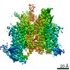 0942MC M: map data used to model this data C: citing same article ( |
|---|---|
| Similar structure data |


-Links
 PDBj
PDBj


- Assembly
| Deposited unit | 
|
|---|---|
| 1 |
|
-Components
| #1: Protein | Mass: 231743.938 Da / Num. of mol.: 1 Source method: isolated from a genetically manipulated source Source: (gene. exp.) Homo sapiens (human) / Gene: SCN5A / Production host: Homo sapiens (human) / References: UniProt: Q14524 | ||||||
|---|---|---|---|---|---|---|---|
| #2: Sugar | ChemComp-NAG /   Type: D-saccharide, beta linking / Mass: 221.208 Da / Num. of mol.: 9 Type: D-saccharide, beta linking / Mass: 221.208 Da / Num. of mol.: 9Source method: isolated from a genetically manipulated source Formula: C8H15NO6 #3: Chemical | ChemComp-QDN / |   Mass: 324.417 Da / Num. of mol.: 1 / Source method: obtained synthetically / Formula: C20H24N2O2 / Comment: antiarrhythmic*YM Mass: 324.417 Da / Num. of mol.: 1 / Source method: obtained synthetically / Formula: C20H24N2O2 / Comment: antiarrhythmic*YMHas ligand of interest | Y | Has protein modification | Y | |
-Experimental details
-Experiment
| Experiment | Method: ELECTRON MICROSCOPY |
|---|---|
| EM experiment | Aggregation state: PARTICLE / 3D reconstruction method: single particle reconstruction |
- Sample preparation
| Component | Name: Voltage-gated sodium channel with drug / Type: COMPLEX / Entity ID: #1 / Source: RECOMBINANT |
|---|---|
| Source (natural) | Organism: Homo sapiens (human) |
| Source (recombinant) | Organism: Homo sapiens (human) |
| Buffer solution | pH: 7.5 |
| Specimen | Embedding applied: NO / Shadowing applied: NO / Staining applied: NO / Vitrification applied: YES |
| Specimen support | Grid material: GOLD / Grid mesh size: 300 divisions/in. / Grid type: Quantifoil R1.2/1.3 |
| Vitrification | Cryogen name: ETHANE |
- Electron microscopy imaging
| Experimental equipment |  Model: Titan Krios / Image courtesy: FEI Company |
|---|---|
| Microscopy | Model: FEI TITAN KRIOS |
| Electron gun | Electron source:  FIELD EMISSION GUN / Accelerating voltage: 300 kV / Illumination mode: FLOOD BEAM FIELD EMISSION GUN / Accelerating voltage: 300 kV / Illumination mode: FLOOD BEAM |
| Electron lens | Mode: BRIGHT FIELD |
| Image recording | Electron dose: 48 e/Å2 / Film or detector model: GATAN K2 SUMMIT (4k x 4k) |
- Processing
| CTF correction | Type: PHASE FLIPPING ONLY |
|---|---|
| 3D reconstruction | Resolution: 3.3 Å / Resolution method: FSC 0.143 CUT-OFF / Num. of particles: 124954 / Symmetry type: POINT |
