 ムービー
ムービー コントローラー


+ データを開く

- 基本情報
基本情報
| 登録情報 | データベース: PDB / ID: 3j2q | |||||||||
|---|---|---|---|---|---|---|---|---|---|---|
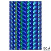
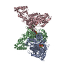
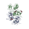
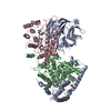
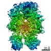
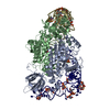
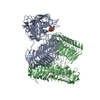
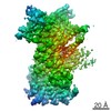
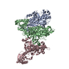
| タイトル | Model of membrane-bound factor VIII organized in 2D crystals | |||||||||
 要素 要素 | (Coagulation factor VIII ...) x 2 | |||||||||
 キーワード キーワード | BLOOD CLOTTING / blood coagulation / cofactor / factor VIII / hemophilia | |||||||||
| 機能・相同性 |  機能・相同性情報 機能・相同性情報Defective F8 accelerates dissociation of the A2 domain / Defective F8 binding to the cell membrane / Defective F8 secretion / Defective F8 sulfation at Y1699 / Gamma carboxylation, hypusinylation, hydroxylation, and arylsulfatase activation / Defective F8 binding to von Willebrand factor / blood coagulation, intrinsic pathway / Cargo concentration in the ER / COPII-coated ER to Golgi transport vesicle / Defective factor IX causes thrombophilia ...Defective F8 accelerates dissociation of the A2 domain / Defective F8 binding to the cell membrane / Defective F8 secretion / Defective F8 sulfation at Y1699 / Gamma carboxylation, hypusinylation, hydroxylation, and arylsulfatase activation / Defective F8 binding to von Willebrand factor / blood coagulation, intrinsic pathway / Cargo concentration in the ER / COPII-coated ER to Golgi transport vesicle / Defective factor IX causes thrombophilia / Defective cofactor function of FVIIIa variant / Defective F9 variant does not activate FX / COPII-mediated vesicle transport / Defective F8 cleavage by thrombin / : / : / endoplasmic reticulum-Golgi intermediate compartment membrane / platelet alpha granule lumen / acute-phase response / Golgi lumen / blood coagulation / Platelet degranulation / oxidoreductase activity / endoplasmic reticulum lumen / copper ion binding / : / extracellular region / plasma membrane 類似検索 - 分子機能 | |||||||||
| 生物種 |  Homo sapiens (ヒト) Homo sapiens (ヒト) | |||||||||
| 手法 | 電子線結晶学 / クライオ電子顕微鏡法 / 解像度: 15 Å | |||||||||
 データ登録者 データ登録者 | Stoilova-Mcphie, S. / Lynch, G.C. / Ludtke, S. / Pettitt, B.M. | |||||||||
 引用 引用 | ジャーナル: Biopolymers / 年: 2013 タイトル: Domain organization of membrane-bound factor VIII. 著者: Svetla Stoilova-McPhie / Gillian C Lynch / Steven Ludtke / B Montgomery Pettitt /  要旨: Factor VIII (FVIII) is the blood coagulation protein which when defective or deficient causes for hemophilia A, a severe hereditary bleeding disorder. Activated FVIII (FVIIIa) is the cofactor to the ...Factor VIII (FVIII) is the blood coagulation protein which when defective or deficient causes for hemophilia A, a severe hereditary bleeding disorder. Activated FVIII (FVIIIa) is the cofactor to the serine protease factor IXa (FIXa) within the membrane-bound Tenase complex, responsible for amplifying its proteolytic activity more than 100,000 times, necessary for normal clot formation. FVIII is composed of two noncovalently linked peptide chains: a light chain (LC) holding the membrane interaction sites and a heavy chain (HC) holding the main FIXa interaction sites. The interplay between the light and heavy chains (HCs) in the membrane-bound state is critical for the biological efficiency of FVIII. Here, we present our cryo-electron microscopy (EM) and structure analysis studies of human FVIII-LC, when helically assembled onto negatively charged single lipid bilayer nanotubes. The resolved FVIII-LC membrane-bound structure supports aspects of our previously proposed FVIII structure from membrane-bound two-dimensional (2D) crystals, such as only the C2 domain interacts directly with the membrane. The LC is oriented differently in the FVIII membrane-bound helical and 2D crystal structures based on EM data, and the existing X-ray structures. This flexibility of the FVIII-LC domain organization in different states is discussed in the light of the FVIIIa-FIXa complex assembly and function. #1: ジャーナル: Blood / 年: 2002 タイトル: 3-Dimensional structure of membrane-bound coagulation factor VIII: modeling of the factor VIII heterodimer within a 3-dimensional density map derived by electron crystallography. 著者: Svetla Stoilova-McPhie / Bruno O Villoutreix / Koen Mertens / Geoffrey Kemball-Cook / Andreas Holzenburg / 要旨: Despite recent studies, the organization of coagulation factor VIII (FVIII) on a phospholipid (PL) membrane is not known in detail. Thus, 2-dimensional (2D) crystals of human FVIII lacking the B ...Despite recent studies, the organization of coagulation factor VIII (FVIII) on a phospholipid (PL) membrane is not known in detail. Thus, 2-dimensional (2D) crystals of human FVIII lacking the B domain were prepared for electron microscopy onto negatively charged PL monolayers. The 3-dimensional (3D) density map of the PL-bound FVIII protein was calculated at 1.5 nm. Existing atomic data and models for FVIII domains were fitted unambiguously within the 3D density map of the molecule. FVIII domains arrangement followed a compact spiral organization with the A3 domains in close association with the C1 and C2 domains near the PL surface. Viewed toward the membrane the A domains' heterotrimer is oriented side-on with the pseudo-3-fold axis almost parallel to the PL surface and A1 fully covering C1. The C2 domain is partially overlapped by the A2 domain of an adjacent molecule in the 2D crystal, favoring close packing. Viewed parallel to the membrane, C2 is slightly inclined to the PL surface covering an area of 12 nm(2). Four C2 loops are embedded within the lipid monolayer at about 0.7 to 1.0 nm depth. C1 forms almost a right angle with C2, its long axis nearly parallel to the membrane. The proposed structure for membrane-bound FVIII results from modeling of the FVIII domains within a 3D density map obtained from electron crystallography and accords with the main biochemical and structural information known to date. A model is proposed for FVIIIa and factor IXa assembly within the membrane-bound factor X-activating complex. (Blood. 2002;99:1215-1223) | |||||||||
| 履歴 |
|
- 構造の表示
構造の表示
| ムービー |
ムービービューア |
|---|---|
| 構造ビューア | 分子: MolmilJmol/JSmol |
- ダウンロードとリンク
-ダウンロード
| PDBx/mmCIF形式 | 3j2q.cif.gz | 284.1 KB | 表示 | PDBx/mmCIF形式 |
|---|---|---|---|---|
| PDB形式 | pdb3j2q.ent.gz | 185.8 KB | 表示 | PDB形式 |
| PDBx/mmJSON形式 | 3j2q.json.gz | ツリー表示 | PDBx/mmJSON形式 | |
| その他 |  その他のダウンロード その他のダウンロード |
-検証レポート
| アーカイブディレクトリ | https://data.pdbj.org/pub/pdb/validation_reports/j2/3j2qftp://data.pdbj.org/pub/pdb/validation_reports/j2/3j2q | HTTPS FTP |
|---|
-関連構造データ


-リンク
 PDBj
PDBj


- 集合体
集合体
| 登録構造単位 | 
| ||||||||
|---|---|---|---|---|---|---|---|---|---|
| 1 |
| ||||||||
| 単位格子 |
|
-要素
-Coagulation factor VIII ... , 2種, 2分子 AB
| #1: タンパク質 | 分子量: 86455.742 Da / 分子数: 1 / 断片: UNP residues 20-764 / 由来タイプ: 組換発現 / 由来: (組換発現) Homo sapiens (ヒト) / 遺伝子: F8, F8C / Cell (発現宿主): ovary発現宿主:   Cricetulus griseus (モンゴルキヌゲネズミ) Cricetulus griseus (モンゴルキヌゲネズミ)参照: UniProt: P00451 |
|---|---|
| #2: タンパク質 | 分子量: 79050.594 Da / 分子数: 1 / 断片: UNP residues 1668-2351 / 由来タイプ: 組換発現 / 由来: (組換発現) Homo sapiens (ヒト) / 遺伝子: F8, F8C / Cell (発現宿主): ovary発現宿主: Cricetulus griseus (モンゴルキヌゲネズミ)参照: UniProt: P00451 |
-糖 , 3種, 4分子 
| #3: 多糖 | 2-acetamido-2-deoxy-beta-D-glucopyranose-(1-4)-2-acetamido-2-deoxy-beta-D-glucopyranose |
|---|---|
| #4: 多糖 | alpha-D-mannopyranose-(1-3)-[beta-D-mannopyranose-(1-6)]beta-D-mannopyranose-(1-4)-2-acetamido-2- ...alpha-D-mannopyranose-(1-3)-[beta-D-mannopyranose-(1-6)]beta-D-mannopyranose-(1-4)-2-acetamido-2-deoxy-beta-D-glucopyranose |
| #7: 糖 |  タイプ: D-saccharide, beta linking / 分子量: 221.208 Da / 分子数: 2 / 由来タイプ: 組換発現 / 式: C8H15NO6 タイプ: D-saccharide, beta linking / 分子量: 221.208 Da / 分子数: 2 / 由来タイプ: 組換発現 / 式: C8H15NO6 |
-非ポリマー , 2種, 3分子 

| #5: 化合物 |  分子量: 63.546 Da / 分子数: 2 / 由来タイプ: 合成 / 式: Cu 分子量: 63.546 Da / 分子数: 2 / 由来タイプ: 合成 / 式: Cu#6: 化合物 | ChemComp-CA / |  分子量: 40.078 Da / 分子数: 1 / 由来タイプ: 合成 / 式: Ca 分子量: 40.078 Da / 分子数: 1 / 由来タイプ: 合成 / 式: Ca |
|---|
-詳細
| Has protein modification | Y |
|---|---|
| 配列の詳細 | THE AUTHORS STATE THAT F1899L IS A NATURAL VARIANT. |
-実験情報
-実験
| 実験 | 手法: 電子線結晶学 |
|---|---|
| EM実験 | 試料の集合状態: 2D ARRAY / 3次元再構成法: 電子線結晶学 |
- 試料調製
試料調製
| 構成要素 | 名称: membrane-bound factor VIII / タイプ: COMPLEX |
|---|---|
| 試料 | 包埋: NO / シャドウイング: NO / 染色: NO / 凍結: YES |
-データ収集
| 顕微鏡 | モデル: JEOL 2010F |
|---|---|
| 電子銃 | 電子線源:  FIELD EMISSION GUN / 加速電圧: 200 kV / 照射モード: FLOOD BEAM FIELD EMISSION GUN / 加速電圧: 200 kV / 照射モード: FLOOD BEAM |
| 電子レンズ | モード: BRIGHT FIELD / 倍率(公称値): 52000 X |
| 撮影 | 電子線照射量: 16 e/Å2 |
| 放射 | プロトコル: SINGLE WAVELENGTH / 単色(M)・ラウエ(L): M / 散乱光タイプ: electron |
| 放射波長 | 相対比: 1 |
- 解析
解析
| 3次元再構成 | 解像度: 15 Å / 対称性のタイプ: 2D CRYSTAL | ||||||||||||
|---|---|---|---|---|---|---|---|---|---|---|---|---|---|
| 精密化ステップ | サイクル: LAST
|
