 Movie
Movie Controller


+ Open data

- Basic information
| Entry | Database: PDB / ID: 3j2s | ||||||
|---|---|---|---|---|---|---|---|
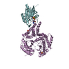
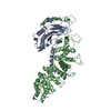
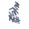
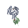
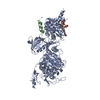
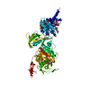
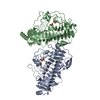
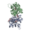
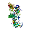
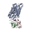
| Title | Membrane-bound factor VIII light chain | ||||||
 Components Components | Coagulation factor VIII light chain | ||||||
 Keywords Keywords | BLOOD CLOTTING / blood coagulation / cofactor / factor VIII / hemophilia / membrane binding | ||||||
| Function / homology |  Function and homology information Function and homology informationDefective F8 accelerates dissociation of the A2 domain / Defective F8 binding to the cell membrane / Defective F8 secretion / Defective F8 sulfation at Y1699 / Gamma carboxylation, hypusinylation, hydroxylation, and arylsulfatase activation / Defective F8 binding to von Willebrand factor / blood coagulation, intrinsic pathway / Cargo concentration in the ER / COPII-coated ER to Golgi transport vesicle / Defective factor IX causes thrombophilia ...Defective F8 accelerates dissociation of the A2 domain / Defective F8 binding to the cell membrane / Defective F8 secretion / Defective F8 sulfation at Y1699 / Gamma carboxylation, hypusinylation, hydroxylation, and arylsulfatase activation / Defective F8 binding to von Willebrand factor / blood coagulation, intrinsic pathway / Cargo concentration in the ER / COPII-coated ER to Golgi transport vesicle / Defective factor IX causes thrombophilia / Defective cofactor function of FVIIIa variant / Defective F9 variant does not activate FX / COPII-mediated vesicle transport / Defective F8 cleavage by thrombin / : / : / endoplasmic reticulum-Golgi intermediate compartment membrane / platelet alpha granule lumen / acute-phase response / Golgi lumen / blood coagulation / Platelet degranulation / oxidoreductase activity / endoplasmic reticulum lumen / copper ion binding / : / extracellular region / plasma membrane Similarity search - Function | ||||||
| Biological species |  Homo sapiens (human) Homo sapiens (human) | ||||||
| Method | ELECTRON MICROSCOPY / helical reconstruction / cryo EM / Resolution: 15 Å | ||||||
 Authors Authors | Stoilova-Mcphie, S. / Lynch, G.C. / Ludtke, S. / Pettitt, B.M. | ||||||
 Citation Citation | Journal: Biopolymers / Year: 2013 Title: Domain organization of membrane-bound factor VIII. Authors: Svetla Stoilova-McPhie / Gillian C Lynch / Steven Ludtke / B Montgomery Pettitt /  Abstract: Factor VIII (FVIII) is the blood coagulation protein which when defective or deficient causes for hemophilia A, a severe hereditary bleeding disorder. Activated FVIII (FVIIIa) is the cofactor to the ...Factor VIII (FVIII) is the blood coagulation protein which when defective or deficient causes for hemophilia A, a severe hereditary bleeding disorder. Activated FVIII (FVIIIa) is the cofactor to the serine protease factor IXa (FIXa) within the membrane-bound Tenase complex, responsible for amplifying its proteolytic activity more than 100,000 times, necessary for normal clot formation. FVIII is composed of two noncovalently linked peptide chains: a light chain (LC) holding the membrane interaction sites and a heavy chain (HC) holding the main FIXa interaction sites. The interplay between the light and heavy chains (HCs) in the membrane-bound state is critical for the biological efficiency of FVIII. Here, we present our cryo-electron microscopy (EM) and structure analysis studies of human FVIII-LC, when helically assembled onto negatively charged single lipid bilayer nanotubes. The resolved FVIII-LC membrane-bound structure supports aspects of our previously proposed FVIII structure from membrane-bound two-dimensional (2D) crystals, such as only the C2 domain interacts directly with the membrane. The LC is oriented differently in the FVIII membrane-bound helical and 2D crystal structures based on EM data, and the existing X-ray structures. This flexibility of the FVIII-LC domain organization in different states is discussed in the light of the FVIIIa-FIXa complex assembly and function. | ||||||
| History |
|
- Structure visualization
| Movie |
Movie viewer |
|---|---|
| Structure viewer | Molecule: MolmilJmol/JSmol |
- Downloads & links
-Download
| PDBx/mmCIF format | 3j2s.cif.gz | 124.7 KB | Display | PDBx/mmCIF format |
|---|---|---|---|---|
| PDB format | pdb3j2s.ent.gz | 91.7 KB | Display | PDB format |
| PDBx/mmJSON format | 3j2s.json.gz | Tree view | PDBx/mmJSON format | |
| Others |  Other downloads Other downloads |
-Validation report
| Arichive directory | https://data.pdbj.org/pub/pdb/validation_reports/j2/3j2sftp://data.pdbj.org/pub/pdb/validation_reports/j2/3j2s | HTTPS FTP |
|---|
-Related structure data
| Related structure data |  5559MC 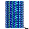 5540C  3j2qC M: map data used to model this data C: citing same article ( |
|---|---|
| Similar structure data |
-Links
 PDBj
PDBj


- Assembly
| Deposited unit | 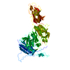
|
|---|---|
| 1 |
|
-Components
| #1: Protein | Mass: 74035.359 Da / Num. of mol.: 1 / Fragment: UNP residues 1710-2356 Source method: isolated from a genetically manipulated source Source: (gene. exp.) Homo sapiens (human) / Gene: F8, F8C / Cell (production host): ovary / Production host:   Cricetulus griseus (Chinese hamster) / References: UniProt: P00451 Cricetulus griseus (Chinese hamster) / References: UniProt: P00451 |
|---|---|
| Has protein modification | Y |
| Sequence details | THE AUTHORS STATE THAT F1899L IS A NATURAL VARIANT. |
-Experimental details
-Experiment
| Experiment | Method: ELECTRON MICROSCOPY |
|---|---|
| EM experiment | Aggregation state: HELICAL ARRAY / 3D reconstruction method: helical reconstruction |
- Sample preparation
| Component |
| ||||||||||||
|---|---|---|---|---|---|---|---|---|---|---|---|---|---|
| Buffer solution | Name: 20 mM Tris-HCl, 150 mM NaCl, 5mM CaCl2 / pH: 7.4 / Details: 20 mM Tris-HCl, 150 mM NaCl, 5mM CaCl2 | ||||||||||||
| Specimen | Conc.: 1 mg/ml / Embedding applied: NO / Shadowing applied: NO / Staining applied: NO / Vitrification applied: YES | ||||||||||||
| Specimen support | Details: 300 mesh R2x2 Quantifoil grids | ||||||||||||
| Vitrification | Instrument: FEI VITROBOT MARK III / Cryogen name: ETHANE / Temp: 95 K / Humidity: 100 % Details: Blot for 3.5 seconds before plunging into liquid ethane (Vitrobot Mark III) Method: Blot for 3.5 seconds before plunging |
- Electron microscopy imaging
| Microscopy | Model: JEOL 2010F / Date: Apr 2, 2010 |
|---|---|
| Electron gun | Electron source:  FIELD EMISSION GUN / Accelerating voltage: 200 kV / Illumination mode: FLOOD BEAM FIELD EMISSION GUN / Accelerating voltage: 200 kV / Illumination mode: FLOOD BEAM |
| Electron lens | Mode: BRIGHT FIELD / Nominal magnification: 52000 X / Calibrated magnification: 52000 X / Nominal defocus max: -4400 nm / Nominal defocus min: -700 nm / Cs: 2 mm |
| Specimen holder | Specimen holder model: GATAN LIQUID NITROGEN / Specimen holder type: GATAN LIQUID NITROGEN / Temperature: 95 K / Tilt angle max: 0 ° / Tilt angle min: 0 ° |
| Image recording | Electron dose: 16 e/Å2 / Film or detector model: GATAN ULTRASCAN 4000 (4k x 4k) / Details: 4096 x 4096 pixels @ 15 microns per pixel |
| Image scans | Num. digital images: 69 |
- Processing
| EM software |
| ||||||||||||||||||||
|---|---|---|---|---|---|---|---|---|---|---|---|---|---|---|---|---|---|---|---|---|---|
| CTF correction | Details: phase corrected based on first Thon ring | ||||||||||||||||||||
| Helical symmerty | Angular rotation/subunit: 10 ° / Axial rise/subunit: 8 Å / Axial symmetry: C1 | ||||||||||||||||||||
| 3D reconstruction | Resolution: 15 Å / Nominal pixel size: 2.9 Å / Actual pixel size: 2.9 Å / Magnification calibration: TMV images / Details: IHRSR combined with SPIDER and EMAN2 / Symmetry type: HELICAL | ||||||||||||||||||||
| Refinement step | Cycle: LAST
|
