Journal: Biopolymers / Year: 2013 Title: Domain organization of membrane-bound factor VIII. Authors: Svetla Stoilova-McPhie / Gillian C Lynch / Steven Ludtke / B Montgomery Pettitt / Abstract: Factor VIII (FVIII) is the blood coagulation protein which when defective or deficient causes for hemophilia A, a severe hereditary bleeding disorder. Activated FVIII (FVIIIa) is the cofactor to the ...Factor VIII (FVIII) is the blood coagulation protein which when defective or deficient causes for hemophilia A, a severe hereditary bleeding disorder. Activated FVIII (FVIIIa) is the cofactor to the serine protease factor IXa (FIXa) within the membrane-bound Tenase complex, responsible for amplifying its proteolytic activity more than 100,000 times, necessary for normal clot formation. FVIII is composed of two noncovalently linked peptide chains: a light chain (LC) holding the membrane interaction sites and a heavy chain (HC) holding the main FIXa interaction sites. The interplay between the light and heavy chains (HCs) in the membrane-bound state is critical for the biological efficiency of FVIII. Here, we present our cryo-electron microscopy (EM) and structure analysis studies of human FVIII-LC, when helically assembled onto negatively charged single lipid bilayer nanotubes. The resolved FVIII-LC membrane-bound structure supports aspects of our previously proposed FVIII structure from membrane-bound two-dimensional (2D) crystals, such as only the C2 domain interacts directly with the membrane. The LC is oriented differently in the FVIII membrane-bound helical and 2D crystal structures based on EM data, and the existing X-ray structures. This flexibility of the FVIII-LC domain organization in different states is discussed in the light of the FVIIIa-FIXa complex assembly and function.
History
Deposition
Dec 12, 2012
-
Header (metadata) release
May 15, 2013
-
Map release
Aug 28, 2013
-
Update
Aug 28, 2013
-
Current status
Aug 28, 2013
Processing site: RCSB / Status: Released
-
Structure visualization
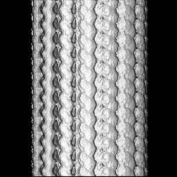
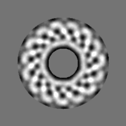
Movie
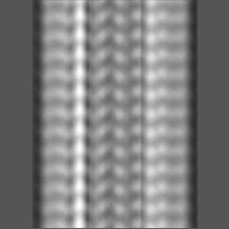
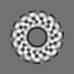
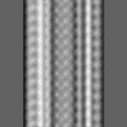
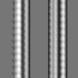
Surface view with section colored by density value
Macromolecule #1: blood coagulation Factor VIII light chain
Macromolecule
Name: blood coagulation Factor VIII light chain / type: protein_or_peptide / ID: 1 / Name.synonym: Hemophilia factor light chain A / Number of copies: 96 / Oligomeric state: helical / Recombinant expression: Yes
Source (natural)
Organism: Homo sapiens (human) / synonym: human / Location in cell: blood plasma
Molecular weight
Experimental: 89 KDa / Theoretical: 90 KDa
Recombinant expression
Organism: Cricetulus griseus (Chinese hamster) / Recombinant cell: CHO
Sequence
UniProtKB: Coagulation factor VIII
-
Experimental details
-
Structure determination
Method
cryo EM
Processing
helical reconstruction
Aggregation state
helical array
-
Sample preparation
Concentration
1 mg/mL
Buffer
pH: 7.4 / Details: 20 mM Tris-HCl 150 mM NaCl, 20 mM EDTA
Grid
Details: 300 mesh R2x2 Quantifoil grids
Vitrification
Cryogen name: ETHANE / Chamber humidity: 100 % / Chamber temperature: 106 K / Instrument: FEI VITROBOT MARK III / Method: Blot for 4.5 seconds before plunging
Details
The protein was mixed in 1:1 w/w ratio with lipid nanotubes solution
-
Electron microscopy
Microscope
JEOL 2010F
Temperature
Min: 90 K / Max: 100 K / Average: 99 K
Alignment procedure
Legacy - Astigmatism: corrected at 400,000 times magnification
Date
Jul 7, 2009
Image recording
Category: CCD / Film or detector model: GATAN ULTRASCAN 4000 (4k x 4k) / Digitization - Sampling interval: 15 µm / Number real images: 69 / Average electron dose: 16 e/Å2 / Details: Each image was acquired for 1 second.
Electron beam
Acceleration voltage: 200 kV / Electron source: FIELD EMISSION GUN
The 2D analysis was performed with EMAN2 and the helical reconstruction with the IHRSR algorithm
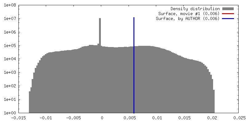
Final reconstruction
Applied symmetry - Helical parameters - Δz: 7.6 Å Applied symmetry - Helical parameters - Δ&Phi: 0.5 ° Algorithm: OTHER / Resolution.type: BY AUTHOR / Resolution: 15.0 Å / Resolution method: OTHER / Software - Name: EMAN2, IHRSR Details: The final 3D reconstructions was calculated from a set of 2043 helical segments cut off from the selected helical tubes at 256 x 256 pixels with 10% overlap.
In the structure databanks used in Yorodumi, some data are registered as the other names, "COVID-19 virus" and "2019-nCoV". Here are the details of the virus and the list of structure data.
Jan 31, 2019. EMDB accession codes are about to change! (news from PDBe EMDB page)
EMDB accession codes are about to change! (news from PDBe EMDB page)
The allocation of 4 digits for EMDB accession codes will soon come to an end. Whilst these codes will remain in use, new EMDB accession codes will include an additional digit and will expand incrementally as the available range of codes is exhausted. The current 4-digit format prefixed with “EMD-” (i.e. EMD-XXXX) will advance to a 5-digit format (i.e. EMD-XXXXX), and so on. It is currently estimated that the 4-digit codes will be depleted around Spring 2019, at which point the 5-digit format will come into force.
The EM Navigator/Yorodumi systems omit the EMD- prefix.
Related info.:Q: What is EMD? / ID/Accession-code notation in Yorodumi/EM Navigator
Yorodumi is a browser for structure data from EMDB, PDB, SASBDB, etc.
This page is also the successor to EM Navigator detail page, and also detail information page/front-end page for Omokage search.
The word "yorodu" (or yorozu) is an old Japanese word meaning "ten thousand". "mi" (miru) is to see.
Related info.:EMDB / PDB / SASBDB / Comparison of 3 databanks / Yorodumi Search / Aug 31, 2016. New EM Navigator & Yorodumi / Yorodumi Papers / Jmol/JSmol / Function and homology information / Changes in new EM Navigator and Yorodumi
 Movie
Movie Controller

 Yorodumi
 Open data
 Basic information
 Map data
 Sample
 Keywords
 Function and homology information
 Homo sapiens (human)
 Authors
 Citation
 Structure visualization
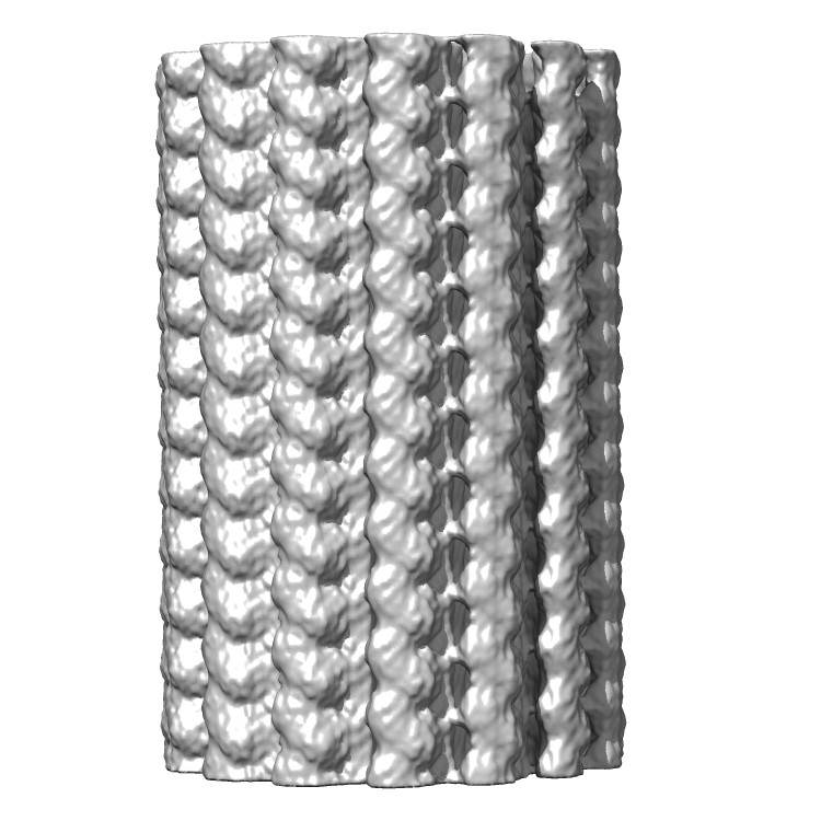
 Downloads & links
 emd_5540_1.jpg
 http://ftp.pdbj.org/pub/emdb/structures/EMD-5540


 Z (Sec.)
 Y (Row.)
 X (Col.)


 Sample components
 Cricetulus griseus (Chinese hamster) / Recombinant cell: CHO
 Processing
 Electron microscopy
 FIELD EMISSION GUN