+ データを開く
- 基本情報
基本情報
| 登録情報 | データベース: PDB / ID: 3j0f | ||||||
|---|---|---|---|---|---|---|---|
| タイトル | Sindbis virion | ||||||
 要素 要素 |
| ||||||
 キーワード キーワード | VIRUS / alphavirus / virus assembly / glycoprotein | ||||||
| 機能・相同性 |  機能・相同性情報 機能・相同性情報icosahedral viral capsid, spike / togavirin / T=4 icosahedral viral capsid / ubiquitin-like protein ligase binding / symbiont-mediated suppression of host toll-like receptor signaling pathway / clathrin-dependent endocytosis of virus by host cell / host cell cytoplasm / membrane fusion / viral translational frameshifting / serine-type endopeptidase activity ...icosahedral viral capsid, spike / togavirin / T=4 icosahedral viral capsid / ubiquitin-like protein ligase binding / symbiont-mediated suppression of host toll-like receptor signaling pathway / clathrin-dependent endocytosis of virus by host cell / host cell cytoplasm / membrane fusion / viral translational frameshifting / serine-type endopeptidase activity / fusion of virus membrane with host endosome membrane / viral envelope / virion attachment to host cell / host cell nucleus / host cell plasma membrane / virion membrane / structural molecule activity / proteolysis / RNA binding / membrane 類似検索 - 分子機能 | ||||||
| 生物種 |  Sindbis virus (シンドビスウイルス) Sindbis virus (シンドビスウイルス) | ||||||
| 手法 | 電子顕微鏡法 / 単粒子再構成法 / クライオ電子顕微鏡法 / 解像度: 7 Å | ||||||
 データ登録者 データ登録者 | Tang, J. / Jose, J. / Zhang, W. / Chipman, P. / Kuhn, R.J. / Baker, T.S. | ||||||
 引用 引用 |  ジャーナル: J Mol Biol / 年: 2011 ジャーナル: J Mol Biol / 年: 2011タイトル: Molecular links between the E2 envelope glycoprotein and nucleocapsid core in Sindbis virus. 著者: Jinghua Tang / Joyce Jose / Paul Chipman / Wei Zhang / Richard J Kuhn / Timothy S Baker /  要旨: A three-dimensional reconstruction of Sindbis virus at 7.0 Å resolution presented here provides a detailed view of the virion structure and includes structural evidence for key interactions that ...A three-dimensional reconstruction of Sindbis virus at 7.0 Å resolution presented here provides a detailed view of the virion structure and includes structural evidence for key interactions that occur between the capsid protein (CP) and transmembrane (TM) glycoproteins E1 and E2. Based on crystal structures of component proteins and homology modeling, we constructed a nearly complete, pseudo-atomic model of the virus. Notably, this includes identification of the 33-residue cytoplasmic domain of E2 (cdE2), which follows a path from the E2 TM helix to the CP where it enters and exits the CP hydrophobic pocket and then folds back to contact the viral membrane. Modeling analysis identified three major contact regions between cdE2 and CP, and the roles of specific residues were probed by molecular genetics. This identified R393 and E395 of cdE2 and Y162 and K252 of CP as critical for virus assembly. The N-termini of the CPs form a contiguous network that interconnects 12 pentameric and 30 hexameric CP capsomers. A single glycoprotein spike cross-links three neighboring CP capsomers as might occur during initiation of virus budding. | ||||||
| 履歴 |
|
- 構造の表示
構造の表示
| ムービー |
 ムービービューア ムービービューア |
|---|---|
| 構造ビューア | 分子:  Molmil Molmil Jmol/JSmol Jmol/JSmol |
- ダウンロードとリンク
- ダウンロード
| PDBx/mmCIF形式 |  3j0f.cif.gz 3j0f.cif.gz | 139.5 KB | 表示 |  PDBx/mmCIF形式 PDBx/mmCIF形式 |
|---|---|---|---|---|
| PDB形式 |  pdb3j0f.ent.gz pdb3j0f.ent.gz | 90.6 KB | 表示 |  PDB形式 PDB形式 |
| PDBx/mmJSON形式 |  3j0f.json.gz 3j0f.json.gz | ツリー表示 |  PDBx/mmJSON形式 PDBx/mmJSON形式 | |
| その他 |  その他のダウンロード その他のダウンロード |
-検証レポート
| 文書・要旨 |  3j0f_validation.pdf.gz 3j0f_validation.pdf.gz | 951.2 KB | 表示 |  wwPDB検証レポート wwPDB検証レポート |
|---|---|---|---|---|
| 文書・詳細版 |  3j0f_full_validation.pdf.gz 3j0f_full_validation.pdf.gz | 952.7 KB | 表示 | |
| XML形式データ |  3j0f_validation.xml.gz 3j0f_validation.xml.gz | 47.1 KB | 表示 | |
| CIF形式データ |  3j0f_validation.cif.gz 3j0f_validation.cif.gz | 74 KB | 表示 | |
| アーカイブディレクトリ |  https://data.pdbj.org/pub/pdb/validation_reports/j0/3j0f https://data.pdbj.org/pub/pdb/validation_reports/j0/3j0f ftp://data.pdbj.org/pub/pdb/validation_reports/j0/3j0f ftp://data.pdbj.org/pub/pdb/validation_reports/j0/3j0f | HTTPS FTP |
-関連構造データ
- リンク
リンク
- 集合体
集合体
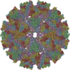
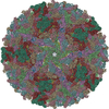
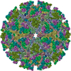
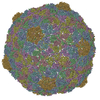
| 登録構造単位 | 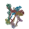
|
|---|---|
| 1 | x 60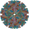
|
| 2 |
|
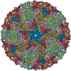
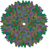
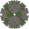
| 3 | x 5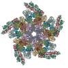
|
| 4 | x 6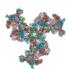
|
| 5 | 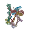
|
| 対称性 | 点対称性: (シェーンフリース記号: I (正20面体型対称)) |
- 要素
要素
| #1: タンパク質 | 分子量: 29410.010 Da / 分子数: 4 / 断片: UNP residues 1-264 / 由来タイプ: 天然 由来: (天然)  Sindbis virus (シンドビスウイルス) Sindbis virus (シンドビスウイルス)参照: UniProt: P03316, togavirin #2: タンパク質 | 分子量: 47416.836 Da / 分子数: 4 / 断片: UNP residues 807-1245 / 由来タイプ: 天然 由来: (天然)  Sindbis virus (シンドビスウイルス) Sindbis virus (シンドビスウイルス)参照: UniProt: P03316 #3: タンパク質 | 分子量: 46982.582 Da / 分子数: 4 / 断片: UNP residues 329-751 / 由来タイプ: 天然 由来: (天然)  Sindbis virus (シンドビスウイルス) Sindbis virus (シンドビスウイルス)参照: UniProt: P03316 |
|---|
-実験情報
-実験
| 実験 | 手法: 電子顕微鏡法 |
|---|---|
| EM実験 | 試料の集合状態: PARTICLE / 3次元再構成法: 単粒子再構成法 |
- 試料調製
試料調製
| 構成要素 | 名称: sindbis virion / タイプ: VIRUS / 詳細: alphavirus |
|---|---|
| ウイルスについての詳細 | 中空か: NO / エンベロープを持つか: YES / ホストのカテゴリ: VERTEBRATES / 単離: STRAIN / タイプ: VIRION |
| 天然宿主 | 生物種: Homo sapiens |
| 試料 | 包埋: NO / シャドウイング: NO / 染色: NO / 凍結: YES |
| 急速凍結 | 凍結剤: ETHANE |
- 電子顕微鏡撮影
| 顕微鏡 | モデル: FEI/PHILIPS CM300FEG/T / 日付: 2001年12月12日 |
|---|---|
| 電子銃 | 電子線源:  FIELD EMISSION GUN / 加速電圧: 300 kV / 照射モード: FLOOD BEAM FIELD EMISSION GUN / 加速電圧: 300 kV / 照射モード: FLOOD BEAM |
| 電子レンズ | モード: BRIGHT FIELD / カメラ長: 0 mm |
| 試料ホルダ | 試料ホルダーモデル: GATAN LIQUID NITROGEN / 資料ホルダタイプ: eucentric / 傾斜角・最大: 0 ° / 傾斜角・最小: 0 ° |
| 撮影 | フィルム・検出器のモデル: KODAK SO-163 FILM |
| 放射 | プロトコル: SINGLE WAVELENGTH / 単色(M)・ラウエ(L): M / 散乱光タイプ: x-ray |
| 放射波長 | 相対比: 1 |
- 解析
解析
| 対称性 | 点対称性: I (正20面体型対称) | ||||||||||||
|---|---|---|---|---|---|---|---|---|---|---|---|---|---|
| 3次元再構成 | 解像度: 7 Å / 対称性のタイプ: POINT | ||||||||||||
| 原子モデル構築 | 空間: REAL | ||||||||||||
| 原子モデル構築 | PDB-ID: 1KXF PDB chain-ID: A / Accession code: 1KXF / Source name: PDB / タイプ: experimental model | ||||||||||||
| 精密化ステップ | サイクル: LAST
|
 ムービー
ムービー コントローラー





 PDBj
PDBj


