ムービー
ムービー コントローラー


+ データを開く

- 基本情報
基本情報
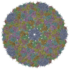
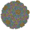
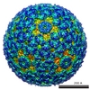
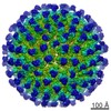
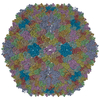
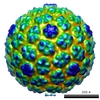
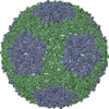
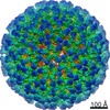
| 登録情報 | データベース: PDB / ID: 3izg | ||||||
|---|---|---|---|---|---|---|---|
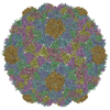
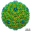
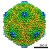
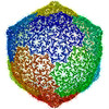
| タイトル | Bacteriophage T7 prohead shell EM-derived atomic model | ||||||
 要素 要素 | Major capsid protein 10A | ||||||
 キーワード キーワード | VIRUS / bacteriophage / capsid maturation / cryoelectron microscopy / morphogenetic intermediate / icosahedral | ||||||
| 機能・相同性 | Capsid Gp10A/Gp10B / : / Major capsid protein / viral capsid / viral translational frameshifting / identical protein binding / Major capsid protein 機能・相同性情報 機能・相同性情報 | ||||||
| 生物種 |   Enterobacteria phage T7 (ファージ) Enterobacteria phage T7 (ファージ) | ||||||
| 手法 | 電子顕微鏡法 / 単粒子再構成法 / クライオ電子顕微鏡法 / 解像度: 10.9 Å | ||||||
 データ登録者 データ登録者 | Ionel, A. / Velazquez-Muriel, J.A. / Agirrezabala, X. / Luque, D. / Cuervo, A. / Caston, J.R. / Valpuesta, J.M. / Martin-Benito, J. / Carrascosa, J.L. | ||||||
 引用 引用 | ジャーナル: J Biol Chem / 年: 2011 タイトル: Molecular rearrangements involved in the capsid shell maturation of bacteriophage T7. 著者: Alina Ionel / Javier A Velázquez-Muriel / Daniel Luque / Ana Cuervo / José R Castón / José M Valpuesta / Jaime Martín-Benito / José L Carrascosa /  要旨: Maturation of dsDNA bacteriophages involves assembling the virus prohead from a limited set of structural components followed by rearrangements required for the stability that is necessary for ...Maturation of dsDNA bacteriophages involves assembling the virus prohead from a limited set of structural components followed by rearrangements required for the stability that is necessary for infecting a host under challenging environmental conditions. Here, we determine the mature capsid structure of T7 at 1 nm resolution by cryo-electron microscopy and compare it with the prohead to reveal the molecular basis of T7 shell maturation. The mature capsid presents an expanded and thinner shell, with a drastic rearrangement of the major protein monomers that increases in their interacting surfaces, in turn resulting in a new bonding lattice. The rearrangements include tilting, in-plane rotation, and radial expansion of the subunits, as well as a relative bending of the A- and P-domains of each subunit. The unique features of this shell transformation, which does not employ the accessory proteins, inserted domains, or molecular interactions observed in other phages, suggest a simple capsid assembling strategy that may have appeared early in the evolution of these viruses. #1: ジャーナル: Structure / 年: 2007タイトル: Quasi-atomic model of bacteriophage t7 procapsid shell: insights into the structure and evolution of a basic fold. 著者: Xabier Agirrezabala / Javier A Velázquez-Muriel / Paulino Gómez-Puertas / Sjors H W Scheres / José M Carazo / José L Carrascosa / 要旨: The existence of similar folds among major structural subunits of viral capsids has shown unexpected evolutionary relationships suggesting common origins irrespective of the capsids' host life domain. ...The existence of similar folds among major structural subunits of viral capsids has shown unexpected evolutionary relationships suggesting common origins irrespective of the capsids' host life domain. Tailed bacteriophages are emerging as one such family, and we have studied the possible existence of the HK97-like fold in bacteriophage T7. The procapsid structure at approximately 10 A resolution was used to obtain a quasi-atomic model by fitting a homology model of the T7 capsid protein gp10 that was based on the atomic structure of the HK97 capsid protein. A number of fold similarities, such as the fitting of domains A and P into the L-shaped procapsid subunit, are evident between both viral systems. A different feature is related to the presence of the amino-terminal domain of gp10 found at the inner surface of the capsid that might play an important role in the interaction of capsid and scaffolding proteins. | ||||||
| 履歴 |
|
- 構造の表示
構造の表示
| ムービー |
ムービービューア |
|---|---|
| 構造ビューア | 分子: MolmilJmol/JSmol |
- ダウンロードとリンク
-ダウンロード
| PDBx/mmCIF形式 | 3izg.cif.gz | 296.7 KB | 表示 | PDBx/mmCIF形式 |
|---|---|---|---|---|
| PDB形式 | pdb3izg.ent.gz | 240.2 KB | 表示 | PDB形式 |
| PDBx/mmJSON形式 | 3izg.json.gz | ツリー表示 | PDBx/mmJSON形式 | |
| その他 |  その他のダウンロード その他のダウンロード |
-検証レポート
| 文書・要旨 | 3izg_validation.pdf.gz | 977.1 KB | 表示 | wwPDB検証レポート |
|---|---|---|---|---|
| 文書・詳細版 | 3izg_full_validation.pdf.gz | 1.1 MB | 表示 | |
| XML形式データ | 3izg_validation.xml.gz | 63.3 KB | 表示 | |
| CIF形式データ | 3izg_validation.cif.gz | 91.6 KB | 表示 | |
| アーカイブディレクトリ | https://data.pdbj.org/pub/pdb/validation_reports/iz/3izgftp://data.pdbj.org/pub/pdb/validation_reports/iz/3izg | HTTPS FTP |
-関連構造データ
-リンク
 PDBj
PDBj- 集合体
集合体
| 登録構造単位 | 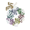
|
|---|---|
| 1 | x 60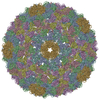
|
| 2 |
|
| 3 | x 5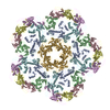
|
| 4 | x 6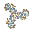
|
| 5 | 
|
| 対称性 | 点対称性: (シェーンフリース記号: I (正20面体型対称)) |
-要素
| #1: タンパク質 | 分子量: 36589.625 Da / 分子数: 7 / 由来タイプ: 天然 / 由来: (天然) Enterobacteria phage T7 (ファージ) / 参照: UniProt: P19726 |
|---|
-実験情報
-実験
| 実験 | 手法: 電子顕微鏡法 |
|---|---|
| EM実験 | 試料の集合状態: PARTICLE / 3次元再構成法: 単粒子再構成法 |
- 試料調製
試料調製
| 構成要素 | 名称: bacteriophage T7 prohead / タイプ: VIRUS / 詳細: gp10A |
|---|---|
| ウイルスについての詳細 | ホストのカテゴリ: BACTERIA / タイプ: VIRION |
| 天然宿主 | 生物種: Escherichia coli |
| 緩衝液 | 名称: 50mM Tris-HCl pH7.7 10mM MgCl2 100mM NaCl / pH: 7.7 / 詳細: 50mM Tris-HCl pH7.7 10mM MgCl2 100mM NaCl |
| 試料 | 包埋: NO / シャドウイング: NO / 染色: NO / 凍結: YES |
| 試料支持 | 詳細: Quantifoil R2/2 carbon grids |
| 急速凍結 | 凍結剤: ETHANE |
- 電子顕微鏡撮影
| 顕微鏡 | モデル: FEI TECNAI 20 |
|---|---|
| 電子銃 | 電子線源:  FIELD EMISSION GUN / 加速電圧: 200 kV / 照射モード: SPOT SCAN FIELD EMISSION GUN / 加速電圧: 200 kV / 照射モード: SPOT SCAN |
| 電子レンズ | モード: BRIGHT FIELD / 倍率(公称値): 50000 X / 倍率(補正後): 51600 X / 最大 デフォーカス(公称値): 1000 nm / 最小 デフォーカス(公称値): 3000 nm / Cs: 2.26 mm |
| 試料ホルダ | 試料ホルダーモデル: GATAN LIQUID NITROGEN / 傾斜角・最大: 0 ° / 傾斜角・最小: 0 ° |
| 撮影 | 電子線照射量: 10 e/Å2 / フィルム・検出器のモデル: KODAK SO-163 FILM |
- 解析
解析
| EMソフトウェア | 名称: SPIDER / カテゴリ: 3次元再構成 | ||||||||||||
|---|---|---|---|---|---|---|---|---|---|---|---|---|---|
| CTF補正 | 詳細: Wiener filter, defocus groups | ||||||||||||
| 対称性 | 点対称性: I (正20面体型対称) | ||||||||||||
| 3次元再構成 | 手法: Spider / 解像度: 10.9 Å / 粒子像の数: 4460 / ピクセルサイズ(実測値): 2.72 Å / 対称性のタイプ: POINT | ||||||||||||
| 原子モデル構築 | PDB-ID: 3E8K Accession code: 3E8K / Source name: PDB / タイプ: experimental model | ||||||||||||
| 精密化ステップ | サイクル: LAST
|