 Movie
Movie Controller

[English] 日本語
 Yorodumi
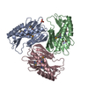
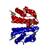
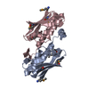
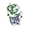
- PDB-3imo: Structure from the mobile metagenome of Vibrio cholerae. Integron... -
+ Open data

- Basic information
| Entry | Database: PDB / ID: 3imo | ||||||
|---|---|---|---|---|---|---|---|
| Title | Structure from the mobile metagenome of Vibrio cholerae. Integron cassette protein VCH_CASS14 | ||||||
 Components Components | Integron cassette protein | ||||||
 Keywords Keywords | UNKNOWN FUNCTION / Novel / Integron protein / Vibrio cholerae / Argentinean O139 strain | ||||||
| Function / homology | Metal Transport, Frataxin; Chain A - #70 / Integron cassette protein VCH_CASS1 chain / Integron cassette protein VCH_CASS1 chain / Metal Transport, Frataxin; Chain A / 2-Layer Sandwich / Alpha Beta / ACETATE ION / Integron cassette protein VCH-CASS1 chain domain-containing protein Function and homology information Function and homology information | ||||||
| Biological species |  Vibrio cholerae O139 (bacteria) Vibrio cholerae O139 (bacteria) | ||||||
| Method |  X-RAY DIFFRACTION / SYNCHROTRON / MOLECULAR REPLACEMENT / Resolution: 1.8 Å X-RAY DIFFRACTION / SYNCHROTRON / MOLECULAR REPLACEMENT / Resolution: 1.8 Å | ||||||
 Authors Authors | Deshpande, C.N. / Sureshan, V. / Harrop, S.J. / Boucher, Y. / Stokes, H.W. / Curmi, P.M.G. / Mabbutt, B.C. | ||||||
 Citation Citation | Journal: Plos One / Year: 2013 Title: Integron gene cassettes: a repository of novel protein folds with distinct interaction sites Authors: Sureshan, V. / Deshpande, C.N. / Boucher, Y. / Koenig, J.E. / Stokes, H.W. / Harrop, S.J. / Curmi, P.M.G. / Mabbutt, B.C. | ||||||
| History |
|
- Structure visualization
| Structure viewer | Molecule: MolmilJmol/JSmol |
|---|
- Downloads & links
-Download
| PDBx/mmCIF format | 3imo.cif.gz | 189.1 KB | Display | PDBx/mmCIF format |
|---|---|---|---|---|
| PDB format | pdb3imo.ent.gz | 152.3 KB | Display | PDB format |
| PDBx/mmJSON format | 3imo.json.gz | Tree view | PDBx/mmJSON format | |
| Others |  Other downloads Other downloads |
-Validation report
| Arichive directory | https://data.pdbj.org/pub/pdb/validation_reports/im/3imoftp://data.pdbj.org/pub/pdb/validation_reports/im/3imo | HTTPS FTP |
|---|
-Related structure data


-Links
 PDBj
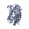
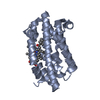
PDBj- Assembly
| Deposited unit | 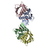
| ||||||||
|---|---|---|---|---|---|---|---|---|---|
| 1 | 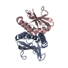
| ||||||||
| 2 | 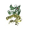
| ||||||||
| Unit cell |
|
-Components
| #1: Protein | Mass: 14742.679 Da / Num. of mol.: 4 Source method: isolated from a genetically manipulated source Source: (gene. exp.) Vibrio cholerae O139 (bacteria) / Strain: Arg3 O139 / Plasmid: pET-15b / Production host: #2: Chemical |   Mass: 59.044 Da / Num. of mol.: 2 / Source method: obtained synthetically / Formula: C2H3O2 Mass: 59.044 Da / Num. of mol.: 2 / Source method: obtained synthetically / Formula: C2H3O2#3: Water | ChemComp-HOH / |  Mass: 18.015 Da / Num. of mol.: 208 / Source method: isolated from a natural source / Formula: H2O Mass: 18.015 Da / Num. of mol.: 208 / Source method: isolated from a natural source / Formula: H2ONonpolymer details | BOUND LIGANDS INCLUDE ACETATE ION FROM CRYSTALLIZ | Sequence details | THERE IS NO UNP REFERENCE SEQUENCE DATABASE FOR THIS PROTEIN AT THE TIME OF PROCESSING. THE FIRST ...THERE IS NO UNP REFERENCE SEQUENCE DATABASE FOR THIS PROTEIN AT THE TIME OF PROCESSING | |
|---|
-Experimental details
-Experiment
| Experiment | Method: X-RAY DIFFRACTION / Number of used crystals: 1 |
|---|
- Sample preparation
| Crystal | Density Matthews: 2.02 Å3/Da / Density % sol: 39.22 % |
|---|---|
| Crystal grow | Temperature: 296 K / Method: vapor diffusion Details: 20% PEG 3350, 0.20M Lithium acetate, Cryoprotectant: Perfluoropolyether PFO-X175/08 (Hampton Research), VAPOR DIFFUSION, temperature 296K |
-Data collection
| Diffraction | Mean temperature: 100 K |
|---|---|
| Diffraction source | Source: SYNCHROTRON / Site: APS  / Beamline: 23-ID-B / Wavelength: 1.03319 Å / Beamline: 23-ID-B / Wavelength: 1.03319 Å |
| Detector | Type: MARMOSAIC 300 mm CCD / Detector: CCD / Date: Aug 2, 2008 |
| Radiation | Protocol: SINGLE WAVELENGTH / Monochromatic (M) / Laue (L): M / Scattering type: x-ray |
| Radiation wavelength | Wavelength: 1.03319 Å / Relative weight: 1 |
| Reflection | Resolution: 1.8→45.08 Å / Num. all: 43702 / Num. obs: 43702 / % possible obs: 99.9 % / Observed criterion σ(F): 0 / Observed criterion σ(I): 0 / Redundancy: 3.8 % / Biso Wilson estimate: 30.693 Å2 / Rmerge(I) obs: 0.066 / Rsym value: 0.077 / Net I/σ(I): 12.3 |
| Reflection shell | Resolution: 1.8→1.9 Å / Redundancy: 3.7 % / Rmerge(I) obs: 0.629 / Mean I/σ(I) obs: 1.8 / Num. unique all: 6365 / Rsym value: 0.737 / % possible all: 100 |
- Processing
| Software |
| |||||||||||||||||||||||||||||||||||||||||||||||||||||||||||||||||||||||||||||||||||||||||||||||||||||||||||||||||||||||||||||||||||||||||||||||||||||||||||||||||||||||||||||||||||||||||||||||||||||||||||||||||||||||||
|---|---|---|---|---|---|---|---|---|---|---|---|---|---|---|---|---|---|---|---|---|---|---|---|---|---|---|---|---|---|---|---|---|---|---|---|---|---|---|---|---|---|---|---|---|---|---|---|---|---|---|---|---|---|---|---|---|---|---|---|---|---|---|---|---|---|---|---|---|---|---|---|---|---|---|---|---|---|---|---|---|---|---|---|---|---|---|---|---|---|---|---|---|---|---|---|---|---|---|---|---|---|---|---|---|---|---|---|---|---|---|---|---|---|---|---|---|---|---|---|---|---|---|---|---|---|---|---|---|---|---|---|---|---|---|---|---|---|---|---|---|---|---|---|---|---|---|---|---|---|---|---|---|---|---|---|---|---|---|---|---|---|---|---|---|---|---|---|---|---|---|---|---|---|---|---|---|---|---|---|---|---|---|---|---|---|---|---|---|---|---|---|---|---|---|---|---|---|---|---|---|---|---|---|---|---|---|---|---|---|---|---|---|---|---|---|---|---|---|
| Refinement | Method to determine structure: MOLECULAR REPLACEMENT Starting model: local model built from low resolution SAD data from p212121 selenomethionine crystals Resolution: 1.8→32.568 Å / Occupancy max: 1 / Occupancy min: 1 / SU ML: 0.26 / Isotropic thermal model: Isotropic / Cross valid method: THROUGHOUT / σ(F): 1.22 / Phase error: 26.02 / Stereochemistry target values: Engh & Huber
| |||||||||||||||||||||||||||||||||||||||||||||||||||||||||||||||||||||||||||||||||||||||||||||||||||||||||||||||||||||||||||||||||||||||||||||||||||||||||||||||||||||||||||||||||||||||||||||||||||||||||||||||||||||||||
| Solvent computation | Shrinkage radii: 0.9 Å / VDW probe radii: 1.11 Å / Solvent model: FLAT BULK SOLVENT MODEL / Bsol: 59.035 Å2 / ksol: 0.391 e/Å3 | |||||||||||||||||||||||||||||||||||||||||||||||||||||||||||||||||||||||||||||||||||||||||||||||||||||||||||||||||||||||||||||||||||||||||||||||||||||||||||||||||||||||||||||||||||||||||||||||||||||||||||||||||||||||||
| Displacement parameters | Biso max: 164.07 Å2 / Biso mean: 45.852 Å2 / Biso min: 17.81 Å2
| |||||||||||||||||||||||||||||||||||||||||||||||||||||||||||||||||||||||||||||||||||||||||||||||||||||||||||||||||||||||||||||||||||||||||||||||||||||||||||||||||||||||||||||||||||||||||||||||||||||||||||||||||||||||||
| Refinement step | Cycle: LAST / Resolution: 1.8→32.568 Å
| |||||||||||||||||||||||||||||||||||||||||||||||||||||||||||||||||||||||||||||||||||||||||||||||||||||||||||||||||||||||||||||||||||||||||||||||||||||||||||||||||||||||||||||||||||||||||||||||||||||||||||||||||||||||||
| Refine LS restraints |
| |||||||||||||||||||||||||||||||||||||||||||||||||||||||||||||||||||||||||||||||||||||||||||||||||||||||||||||||||||||||||||||||||||||||||||||||||||||||||||||||||||||||||||||||||||||||||||||||||||||||||||||||||||||||||
| LS refinement shell |
| |||||||||||||||||||||||||||||||||||||||||||||||||||||||||||||||||||||||||||||||||||||||||||||||||||||||||||||||||||||||||||||||||||||||||||||||||||||||||||||||||||||||||||||||||||||||||||||||||||||||||||||||||||||||||
| Refinement TLS params. | Method: refined / Refine-ID: X-RAY DIFFRACTION
| |||||||||||||||||||||||||||||||||||||||||||||||||||||||||||||||||||||||||||||||||||||||||||||||||||||||||||||||||||||||||||||||||||||||||||||||||||||||||||||||||||||||||||||||||||||||||||||||||||||||||||||||||||||||||
| Refinement TLS group |
|
