 Movie
Movie Controller

[English] 日本語
 Yorodumi
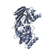
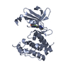
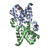
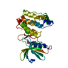
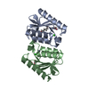
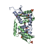
- PDB-3fy6: Structure from the mobile metagenome of V. Cholerae. Integron cas... -
+ Open data

- Basic information
| Entry | Database: PDB / ID: 3fy6 | ||||||
|---|---|---|---|---|---|---|---|
| Title | Structure from the mobile metagenome of V. Cholerae. Integron cassette protein VCH_CASS3 | ||||||
 Components Components | Integron cassette protein | ||||||
 Keywords Keywords | STRUCTURAL GENOMICS / UNKNOWN FUNCTION / Novel / Integron cassette protein / Vibrio cholerae / Oyster pond / Woodshole / USA / PSI-2 / Protein Structure Initiative / Midwest Center for Structural Genomics / MCSG | ||||||
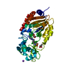
| Function / homology | Integron cassette protein fold / Integron cassette protein superfamily / : / : / Integron cassette protein / 2-Layer Sandwich / Alpha Beta / Integron cassette protein Function and homology information Function and homology information | ||||||
| Biological species |   Vibrio cholerae (bacteria) Vibrio cholerae (bacteria) | ||||||
| Method |  X-RAY DIFFRACTION / SYNCHROTRON / SAD / Resolution: 2.1 Å X-RAY DIFFRACTION / SYNCHROTRON / SAD / Resolution: 2.1 Å | ||||||
 Authors Authors | Deshpande, C.N. / Sureshan, V. / Harrop, S.J. / Boucher, Y. / Xu, X. / Cui, H. / Edwards, A. / Savchenko, A. / Joachimiak, A. / Tan, K. ...Deshpande, C.N. / Sureshan, V. / Harrop, S.J. / Boucher, Y. / Xu, X. / Cui, H. / Edwards, A. / Savchenko, A. / Joachimiak, A. / Tan, K. / Stokes, H.W. / Curmi, P.M.G. / Mabbutt, B.C. / Midwest Center for Structural Genomics (MCSG) | ||||||
 Citation Citation | Journal: Plos One / Year: 2013 Title: Integron gene cassettes: a repository of novel protein folds with distinct interaction sites. Authors: Sureshan, V. / Deshpande, C.N. / Boucher, Y. / Koenig, J.E. / Stokes, H.W. / Harrop, S.J. / Curmi, P.M. / Mabbutt, B.C. | ||||||
| History |
|
- Structure visualization
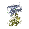
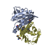
| Structure viewer | Molecule: MolmilJmol/JSmol |
|---|
- Downloads & links
-Download
| PDBx/mmCIF format | 3fy6.cif.gz | 215.9 KB | Display | PDBx/mmCIF format |
|---|---|---|---|---|
| PDB format | pdb3fy6.ent.gz | 174.9 KB | Display | PDB format |
| PDBx/mmJSON format | 3fy6.json.gz | Tree view | PDBx/mmJSON format | |
| Others |  Other downloads Other downloads |
-Validation report
| Arichive directory | https://data.pdbj.org/pub/pdb/validation_reports/fy/3fy6ftp://data.pdbj.org/pub/pdb/validation_reports/fy/3fy6 | HTTPS FTP |
|---|
-Related structure data
| Related structure data | 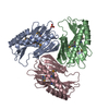 3fuyC 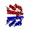 3fxhC  3if4C 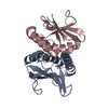 3imoC  3jrtC C: citing same article ( |
|---|---|
| Similar structure data | |
| Other databases |
-Links
 PDBj
PDBj- Assembly
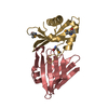
| Deposited unit | 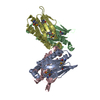
| ||||||||
|---|---|---|---|---|---|---|---|---|---|
| 1 | 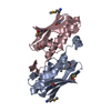
| ||||||||
| 2 | 
| ||||||||
| Unit cell |
|
-Components
| #1: Protein | Mass: 15325.311 Da / Num. of mol.: 4 Source method: isolated from a genetically manipulated source Source: (gene. exp.) Vibrio cholerae (bacteria) / Strain: OPVCH_OP8A / Plasmid: p15TV-L / Production host: #2: Water | ChemComp-HOH / |  Mass: 18.015 Da / Num. of mol.: 323 / Source method: isolated from a natural source / Formula: H2O Mass: 18.015 Da / Num. of mol.: 323 / Source method: isolated from a natural source / Formula: H2OHas protein modification | Y | |
|---|
-Experimental details
-Experiment
| Experiment | Method: X-RAY DIFFRACTION / Number of used crystals: 2 |
|---|
- Sample preparation
| Crystal | Density Matthews: 2.31 Å3/Da / Density % sol: 46.68 % |
|---|---|
| Crystal grow | Method: vapor diffusion / pH: 4.6 Details: 0.1M sodium acetate , 2.0M sodium Formate, pH 4.6, VAPOR DIFFUSION |
-Data collection
| Diffraction | Mean temperature: 100 K |
|---|---|
| Diffraction source | Source: SYNCHROTRON / Site: APS  / Beamline: 19-ID / Wavelength: 0.97929 Å / Beamline: 19-ID / Wavelength: 0.97929 Å |
| Detector | Type: ADSC QUANTUM 315 / Detector: CCD / Date: Oct 24, 2008 |
| Radiation | Protocol: SINGLE WAVELENGTH / Monochromatic (M) / Laue (L): M / Scattering type: x-ray |
| Radiation wavelength | Wavelength: 0.97929 Å / Relative weight: 1 |
| Reflection | Resolution: 2.1→74.43 Å / Num. all: 33584 / Num. obs: 33584 / % possible obs: 99.3 % / Observed criterion σ(F): 0 / Observed criterion σ(I): 0 / Redundancy: 3.9 % / Biso Wilson estimate: 24.992 Å2 / Rmerge(I) obs: 0.088 / Rsym value: 0.13 / Net I/σ(I): 9.4 |
| Reflection shell | Resolution: 2.1→2.21 Å / Redundancy: 3.9 % / Rmerge(I) obs: 0.403 / Mean I/σ(I) obs: 3 / Rsym value: 0.508 / % possible all: 99.2 |
- Processing
| Software |
| ||||||||||||||||||||||||||||||||||||||||||||||||||||||||||||||||||||||||||||||||||||||||||||||||||||||||||||||||||||||||||||||||||
|---|---|---|---|---|---|---|---|---|---|---|---|---|---|---|---|---|---|---|---|---|---|---|---|---|---|---|---|---|---|---|---|---|---|---|---|---|---|---|---|---|---|---|---|---|---|---|---|---|---|---|---|---|---|---|---|---|---|---|---|---|---|---|---|---|---|---|---|---|---|---|---|---|---|---|---|---|---|---|---|---|---|---|---|---|---|---|---|---|---|---|---|---|---|---|---|---|---|---|---|---|---|---|---|---|---|---|---|---|---|---|---|---|---|---|---|---|---|---|---|---|---|---|---|---|---|---|---|---|---|---|---|
| Refinement | Method to determine structure: SAD / Resolution: 2.1→44.867 Å / SU ML: 0.26 / Isotropic thermal model: Isotropic / σ(F): 0.94 / Phase error: 20.65 / Stereochemistry target values: MLHL
| ||||||||||||||||||||||||||||||||||||||||||||||||||||||||||||||||||||||||||||||||||||||||||||||||||||||||||||||||||||||||||||||||||
| Solvent computation | Shrinkage radii: 0.9 Å / VDW probe radii: 1.11 Å / Solvent model: FLAT BULK SOLVENT MODEL / Bsol: 44.126 Å2 / ksol: 0.389 e/Å3 | ||||||||||||||||||||||||||||||||||||||||||||||||||||||||||||||||||||||||||||||||||||||||||||||||||||||||||||||||||||||||||||||||||
| Refinement step | Cycle: LAST / Resolution: 2.1→44.867 Å
| ||||||||||||||||||||||||||||||||||||||||||||||||||||||||||||||||||||||||||||||||||||||||||||||||||||||||||||||||||||||||||||||||||
| Refine LS restraints |
| ||||||||||||||||||||||||||||||||||||||||||||||||||||||||||||||||||||||||||||||||||||||||||||||||||||||||||||||||||||||||||||||||||
| LS refinement shell |
| ||||||||||||||||||||||||||||||||||||||||||||||||||||||||||||||||||||||||||||||||||||||||||||||||||||||||||||||||||||||||||||||||||
| Refinement TLS params. | Refine-ID: X-RAY DIFFRACTION
| ||||||||||||||||||||||||||||||||||||||||||||||||||||||||||||||||||||||||||||||||||||||||||||||||||||||||||||||||||||||||||||||||||
| Refinement TLS group | Selection details: chain D |
