 Movie
Movie Controller

[English] 日本語
 Yorodumi
- PDB-1vj2: Crystal structure of a novel family of manganese-containing cupin... -
+ Open data

- Basic information
| Entry | Database: PDB / ID: 1vj2 | |||||||||
|---|---|---|---|---|---|---|---|---|---|---|
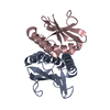
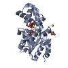
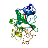
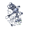
| Title | Crystal structure of a novel family of manganese-containing cupin (tm1459) from thermotoga maritima at 1.65 A resolution | |||||||||
 Components Components | novel manganese-containing cupin TM1459 | |||||||||
 Keywords Keywords | METAL BINDING PROTEIN / Novel manganese-containing cupin / structural genomics / Joint Center for Structural Genomics / JCSG / Protein Structure Initiative / PSI | |||||||||
| Function / homology |  Function and homology information Function and homology information | |||||||||
| Biological species |   Thermotoga maritima (bacteria) Thermotoga maritima (bacteria) | |||||||||
| Method |  X-RAY DIFFRACTION / SYNCHROTRON / MOLECULAR REPLACEMENT / Resolution: 1.65 Å X-RAY DIFFRACTION / SYNCHROTRON / MOLECULAR REPLACEMENT / Resolution: 1.65 Å | |||||||||
 Authors Authors | Joint Center for Structural Genomics (JCSG) | |||||||||
 Citation Citation | Journal: Proteins / Year: 2004 Title: Crystal structure of a novel manganese-containing cupin (TM1459) from Thermotoga maritima at 1.65 A resolution. Authors: Jaroszewski, L. / Schwarzenbacher, R. / von Delft, F. / McMullan, D. / Brinen, L.S. / Canaves, J.M. / Dai, X. / Deacon, A.M. / DiDonato, M. / Elsliger, M.A. / Eshagi, S. / Floyd, R. / ...Authors: Jaroszewski, L. / Schwarzenbacher, R. / von Delft, F. / McMullan, D. / Brinen, L.S. / Canaves, J.M. / Dai, X. / Deacon, A.M. / DiDonato, M. / Elsliger, M.A. / Eshagi, S. / Floyd, R. / Godzik, A. / Grittini, C. / Grzechnik, S.K. / Hampton, E. / Levin, I. / Karlak, C. / Klock, H.E. / Koesema, E. / Kovarik, J.S. / Kreusch, A. / Kuhn, P. / Lesley, S.A. / McPhillips, T.M. / Miller, M.D. / Morse, A. / Moy, K. / Ouyang, J. / Page, R. / Quijano, K. / Reyes, R. / Rezezadeh, F. / Robb, A. / Sims, E. / Spraggon, G. / Stevens, R.C. / van den Bedem, H. / Velasquez, J. / Vincent, J. / Wang, X. / West, B. / Wolf, G. / Xu, Q. / Hodgson, K.O. / Wooley, J. / Wilson, I.A. | |||||||||
| History |
| |||||||||
| Remark 600 | HETEROGEN THE ACTIVE SITE OF SUBUNIT A NEAR CYS106 HAS BEEN MODELED AS TYPE UNL. |
- Structure visualization
| Structure viewer | Molecule: MolmilJmol/JSmol |
|---|
- Downloads & links
-Download
| PDBx/mmCIF format | 1vj2.cif.gz | 65.7 KB | Display | PDBx/mmCIF format |
|---|---|---|---|---|
| PDB format | pdb1vj2.ent.gz | 47.1 KB | Display | PDB format |
| PDBx/mmJSON format | 1vj2.json.gz | Tree view | PDBx/mmJSON format | |
| Others |  Other downloads Other downloads |
-Validation report
| Arichive directory | https://data.pdbj.org/pub/pdb/validation_reports/vj/1vj2ftp://data.pdbj.org/pub/pdb/validation_reports/vj/1vj2 | HTTPS FTP |
|---|
-Related structure data
| Related structure data |  1fi2S S: Starting model for refinement |
|---|---|
| Similar structure data | |
| Other databases |


-Links
 PDBj
PDBj

- Assembly
| Deposited unit | 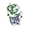
| ||||||||
|---|---|---|---|---|---|---|---|---|---|
| 1 |
| ||||||||
| Unit cell |
|
-Components
| #1: Protein | Mass: 14590.701 Da / Num. of mol.: 2 Source method: isolated from a genetically manipulated source Source: (gene. exp.) Thermotoga maritima (bacteria) / Gene: TM1459 / Production host: #2: Chemical |   Mass: 54.938 Da / Num. of mol.: 2 / Source method: obtained synthetically / Formula: Mn Mass: 54.938 Da / Num. of mol.: 2 / Source method: obtained synthetically / Formula: Mn#3: Chemical | ChemComp-UNL / | Num. of mol.: 1 / Source method: obtained synthetically #4: Water | ChemComp-HOH / |  Mass: 18.015 Da / Num. of mol.: 169 / Source method: isolated from a natural source / Formula: H2O Mass: 18.015 Da / Num. of mol.: 169 / Source method: isolated from a natural source / Formula: H2O |
|---|
-Experimental details
-Experiment
| Experiment | Method: X-RAY DIFFRACTION / Number of used crystals: 1 |
|---|
- Sample preparation
| Crystal | Density Matthews: 2.71 Å3/Da / Density % sol: 54.2 % | ||||||||||||||||||||||||||||||||||||||||||||||||||||||||
|---|---|---|---|---|---|---|---|---|---|---|---|---|---|---|---|---|---|---|---|---|---|---|---|---|---|---|---|---|---|---|---|---|---|---|---|---|---|---|---|---|---|---|---|---|---|---|---|---|---|---|---|---|---|---|---|---|---|
| Crystal grow | Temperature: 293 K / Method: vapor diffusion, sitting drop, nanodrop / pH: 5.2 Details: 50% (v/v) PEG-200, 0.1M Phosphate-citrate pH 4.2 0.2M NaCl, pH 5.2, VAPOR DIFFUSION,SITTING DROP,NANODROP, temperature 293K | ||||||||||||||||||||||||||||||||||||||||||||||||||||||||
| Crystal grow | *PLUS pH: 7.9 / Method: vapor diffusion | ||||||||||||||||||||||||||||||||||||||||||||||||||||||||
| Components of the solutions | *PLUS
|
-Data collection
| Diffraction | Mean temperature: 100 K |
|---|---|
| Diffraction source | Source: SYNCHROTRON / Site: SSRL  / Beamline: BL11-1 / Wavelength: 0.972386 / Beamline: BL11-1 / Wavelength: 0.972386 |
| Detector | Type: ADSC QUANTUM 315 / Detector: CCD / Date: Jan 9, 2002 / Details: flat mirror |
| Radiation | Monochromator: single crystal Si(311) bent monochromator / Protocol: SINGLE WAVELENGTH / Monochromatic (M) / Laue (L): M / Scattering type: x-ray |
| Radiation wavelength | Wavelength: 0.972386 Å / Relative weight: 1 |
| Reflection | Resolution: 1.65→48.13 Å / Num. all: 35029 / Num. obs: 35029 / % possible obs: 97.8 % / Redundancy: 3.6 % / Biso Wilson estimate: 30.49 Å2 / Rsym value: 0.057 / Net I/σ(I): 15.6 |
| Reflection shell | Resolution: 1.65→1.74 Å / Redundancy: 2.7 % / Mean I/σ(I) obs: 1.9 / Num. unique all: 4592 / Rsym value: 0.466 / % possible all: 87.6 |
| Reflection | *PLUS Highest resolution: 1.65 Å / Num. measured all: 124395 / Rmerge(I) obs: 0.067 |
| Reflection shell | *PLUS % possible obs: 87.6 % / Rmerge(I) obs: 0.577 |
- Processing
| Software |
| |||||||||||||||||||||||||||||||||||||||||||||||||||||||||||||||||||||||||||||||||||||||||||||||||||||||||||||||||||
|---|---|---|---|---|---|---|---|---|---|---|---|---|---|---|---|---|---|---|---|---|---|---|---|---|---|---|---|---|---|---|---|---|---|---|---|---|---|---|---|---|---|---|---|---|---|---|---|---|---|---|---|---|---|---|---|---|---|---|---|---|---|---|---|---|---|---|---|---|---|---|---|---|---|---|---|---|---|---|---|---|---|---|---|---|---|---|---|---|---|---|---|---|---|---|---|---|---|---|---|---|---|---|---|---|---|---|---|---|---|---|---|---|---|---|---|---|
| Refinement | Method to determine structure: MOLECULAR REPLACEMENT Starting model: 1FI2 Resolution: 1.65→33.07 Å / Cor.coef. Fo:Fc: 0.953 / Cor.coef. Fo:Fc free: 0.937 / SU B: 6.097 / SU ML: 0.097 / Isotropic thermal model: ISOTROPIC / Cross valid method: THROUGHOUT / ESU R: 0.1 / ESU R Free: 0.104 / Stereochemistry target values: MAXIMUM LIKELIHOOD Details: 1. HYDROGENS HAVE BEEN ADDED IN THE RIDING POSITIONS. 2. THE BOUND METAL WAS IDENTIFIED AS TRANSITION METAL BY ITS COORDINATION GEOMETRY, AND AS MANGANESE BY COMPARISON WITH THE HOMOLOGOUS ...Details: 1. HYDROGENS HAVE BEEN ADDED IN THE RIDING POSITIONS. 2. THE BOUND METAL WAS IDENTIFIED AS TRANSITION METAL BY ITS COORDINATION GEOMETRY, AND AS MANGANESE BY COMPARISON WITH THE HOMOLOGOUS STRUCTURE TM1287 (1O4T) AS WELL AS BY HAVING BEST B-FACTOR AGREEMENT WITH SURROUNDING ATOMS. 3. THE ACTIVE SITE OF SUBUNIT A NEAR CYS106 HAS BEEN MODELED AS TYPE UNL.
| |||||||||||||||||||||||||||||||||||||||||||||||||||||||||||||||||||||||||||||||||||||||||||||||||||||||||||||||||||
| Solvent computation | Ion probe radii: 0.8 Å / Shrinkage radii: 0.8 Å / VDW probe radii: 1.2 Å / Solvent model: BABINET MODEL WITH MASK | |||||||||||||||||||||||||||||||||||||||||||||||||||||||||||||||||||||||||||||||||||||||||||||||||||||||||||||||||||
| Displacement parameters | Biso mean: 24.615 Å2
| |||||||||||||||||||||||||||||||||||||||||||||||||||||||||||||||||||||||||||||||||||||||||||||||||||||||||||||||||||
| Refinement step | Cycle: LAST / Resolution: 1.65→33.07 Å
| |||||||||||||||||||||||||||||||||||||||||||||||||||||||||||||||||||||||||||||||||||||||||||||||||||||||||||||||||||
| Refine LS restraints |
| |||||||||||||||||||||||||||||||||||||||||||||||||||||||||||||||||||||||||||||||||||||||||||||||||||||||||||||||||||
| LS refinement shell | Resolution: 1.65→1.693 Å / Total num. of bins used: 20
| |||||||||||||||||||||||||||||||||||||||||||||||||||||||||||||||||||||||||||||||||||||||||||||||||||||||||||||||||||
| Refinement TLS params. | Method: refined / Refine-ID: X-RAY DIFFRACTION
| |||||||||||||||||||||||||||||||||||||||||||||||||||||||||||||||||||||||||||||||||||||||||||||||||||||||||||||||||||
| Refinement TLS group | Refine-ID: X-RAY DIFFRACTION / Selection: ALL
| |||||||||||||||||||||||||||||||||||||||||||||||||||||||||||||||||||||||||||||||||||||||||||||||||||||||||||||||||||
| Software | *PLUS Name: REFMAC / Version: 5.1.9999 / Classification: refinement | |||||||||||||||||||||||||||||||||||||||||||||||||||||||||||||||||||||||||||||||||||||||||||||||||||||||||||||||||||
| Refinement | *PLUS Rfactor Rfree: 0.249 / Rfactor Rwork: 0.208 | |||||||||||||||||||||||||||||||||||||||||||||||||||||||||||||||||||||||||||||||||||||||||||||||||||||||||||||||||||
| Solvent computation | *PLUS | |||||||||||||||||||||||||||||||||||||||||||||||||||||||||||||||||||||||||||||||||||||||||||||||||||||||||||||||||||
| Displacement parameters | *PLUS | |||||||||||||||||||||||||||||||||||||||||||||||||||||||||||||||||||||||||||||||||||||||||||||||||||||||||||||||||||
| Refine LS restraints | *PLUS
|
