 Movie
Movie Controller


+ Open data

- Basic information
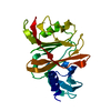
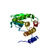
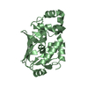
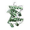
| Entry | Database: PDB / ID: 6rvu | |||||||||
|---|---|---|---|---|---|---|---|---|---|---|
| Title | Crystal structure of the Burkholderia Lethal Factor 1 (BLF1) | |||||||||
 Components Components | Lethal Factor 1 (BLF1) | |||||||||
 Keywords Keywords | TOXIN / Glutamine deamidase toxin / cysteine protease / CNF1 family | |||||||||
| Function / homology | Burkholderia lethal factor 1 / Burkholderia lethal factor 1 / symbiont-mediated suppression of host translation initiation / Uncharacterized protein Function and homology information Function and homology information | |||||||||
| Biological species |  Burkholderia pseudomallei (bacteria) Burkholderia pseudomallei (bacteria) | |||||||||
| Method |  X-RAY DIFFRACTION / SYNCHROTRON / MOLECULAR REPLACEMENT / molecular replacement / Resolution: 0.99 Å X-RAY DIFFRACTION / SYNCHROTRON / MOLECULAR REPLACEMENT / molecular replacement / Resolution: 0.99 Å | |||||||||
 Authors Authors | Mobbs, G.W. / Aziz, A.A. / Blackburn, G.M. / Sedelnikova, S.E. / Minshull, T.C. / Dickman, M.J. / Baker, P.J. / Nathan, S. / Firdaus-Raih, M. / Rice, D.W. | |||||||||
| Funding support |  United Kingdom, 2items United Kingdom, 2items
| |||||||||
 Citation Citation | Journal: Commun Biol / Year: 2022 Title: Molecular basis of specificity and deamidation of eIF4A by Burkholderia Lethal Factor 1. Authors: Mobbs, G.W. / Aziz, A.A. / Dix, S.R. / Blackburn, G.M. / Sedelnikova, S.E. / Minshull, T.C. / Dickman, M.J. / Baker, P.J. / Nathan, S. / Raih, M.F. / Rice, D.W. | |||||||||
| History |
|
- Structure visualization
| Structure viewer | Molecule: MolmilJmol/JSmol |
|---|
- Downloads & links
-Download
| PDBx/mmCIF format | 6rvu.cif.gz | 116.9 KB | Display | PDBx/mmCIF format |
|---|---|---|---|---|
| PDB format | pdb6rvu.ent.gz | 88.8 KB | Display | PDB format |
| PDBx/mmJSON format | 6rvu.json.gz | Tree view | PDBx/mmJSON format | |
| Others |  Other downloads Other downloads |
-Validation report
| Arichive directory | https://data.pdbj.org/pub/pdb/validation_reports/rv/6rvuftp://data.pdbj.org/pub/pdb/validation_reports/rv/6rvu | HTTPS FTP |
|---|
-Related structure data
| Related structure data |  7ppzC  7pq0C 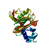 3tu8S  6rrz 6rvt S: Starting model for refinement C: citing same article ( |
|---|---|
| Similar structure data |


-Links
 PDBj
PDBj- Assembly
| Deposited unit | 
| ||||||||
|---|---|---|---|---|---|---|---|---|---|
| 1 |
| ||||||||
| Unit cell |
|
-Components
| #1: Protein | Mass: 25531.344 Da / Num. of mol.: 1 Source method: isolated from a genetically manipulated source Source: (gene. exp.) Burkholderia pseudomallei (strain K96243) (bacteria)Strain: K96243 / Gene: BPSL1549 / Production host: | ||
|---|---|---|---|
| #2: Chemical | ChemComp-EDO /   Mass: 62.068 Da / Num. of mol.: 4 / Source method: obtained synthetically / Formula: C2H6O2 Mass: 62.068 Da / Num. of mol.: 4 / Source method: obtained synthetically / Formula: C2H6O2#3: Water | ChemComp-HOH / |  Mass: 18.015 Da / Num. of mol.: 369 / Source method: isolated from a natural source / Formula: H2O Mass: 18.015 Da / Num. of mol.: 369 / Source method: isolated from a natural source / Formula: H2O |
-Experimental details
-Experiment
| Experiment | Method: X-RAY DIFFRACTION / Number of used crystals: 1 |
|---|
- Sample preparation
| Crystal | Density Matthews: 2.09 Å3/Da / Density % sol: 41.14 % |
|---|---|
| Crystal grow | Temperature: 290 K / Method: vapor diffusion, sitting drop / pH: 7.5 / Details: 0.2 M potassium thiocyanate, 20 % (w/v) PEG 3350 |
-Data collection
| Diffraction | Mean temperature: 100 K / Serial crystal experiment: N | ||||||||||||||||||||||||||||||
|---|---|---|---|---|---|---|---|---|---|---|---|---|---|---|---|---|---|---|---|---|---|---|---|---|---|---|---|---|---|---|---|
| Diffraction source | Source: SYNCHROTRON / Site: Diamond / Beamline: I03 / Wavelength: 0.9763 Å | ||||||||||||||||||||||||||||||
| Detector | Type: DECTRIS PILATUS 6M-F / Detector: PIXEL / Date: Dec 17, 2017 | ||||||||||||||||||||||||||||||
| Radiation | Protocol: SINGLE WAVELENGTH / Monochromatic (M) / Laue (L): M / Scattering type: x-ray | ||||||||||||||||||||||||||||||
| Radiation wavelength | Wavelength: 0.9763 Å / Relative weight: 1 | ||||||||||||||||||||||||||||||
| Reflection | Resolution: 0.99→44.58 Å / Num. obs: 93887 / % possible obs: 86.7 % / Redundancy: 5.8 % / CC1/2: 0.996 / Rmerge(I) obs: 0.078 / Rpim(I) all: 0.032 / Rrim(I) all: 0.084 / Net I/σ(I): 12.6 / Num. measured all: 548111 / Scaling rejects: 651 | ||||||||||||||||||||||||||||||
| Reflection shell | Diffraction-ID: 1
|
-Phasing
| Phasing | Method: molecular replacement |
|---|
- Processing
| Software |
| |||||||||||||||||||||||||||||||||||||||||||||||||||||||||||||||||
|---|---|---|---|---|---|---|---|---|---|---|---|---|---|---|---|---|---|---|---|---|---|---|---|---|---|---|---|---|---|---|---|---|---|---|---|---|---|---|---|---|---|---|---|---|---|---|---|---|---|---|---|---|---|---|---|---|---|---|---|---|---|---|---|---|---|---|
| Refinement | Method to determine structure: MOLECULAR REPLACEMENT Starting model: 3TU8 Resolution: 0.99→41.69 Å / Cor.coef. Fo:Fc: 0.981 / Cor.coef. Fo:Fc free: 0.977 / SU B: 0.598 / SU ML: 0.014 / Cross valid method: THROUGHOUT / σ(F): 0 / ESU R: 0.023 / ESU R Free: 0.024 Details: HYDROGENS HAVE BEEN ADDED IN THE RIDING POSITIONS U VALUES : REFINED INDIVIDUALLY
| |||||||||||||||||||||||||||||||||||||||||||||||||||||||||||||||||
| Solvent computation | Ion probe radii: 0.8 Å / Shrinkage radii: 0.8 Å / VDW probe radii: 1.2 Å | |||||||||||||||||||||||||||||||||||||||||||||||||||||||||||||||||
| Displacement parameters | Biso max: 133.97 Å2 / Biso mean: 13.399 Å2 / Biso min: 5.08 Å2
| |||||||||||||||||||||||||||||||||||||||||||||||||||||||||||||||||
| Refinement step | Cycle: final / Resolution: 0.99→41.69 Å
| |||||||||||||||||||||||||||||||||||||||||||||||||||||||||||||||||
| Refine LS restraints |
| |||||||||||||||||||||||||||||||||||||||||||||||||||||||||||||||||
| LS refinement shell | Resolution: 0.994→1.019 Å / Rfactor Rfree error: 0 / Total num. of bins used: 20
|
