Resolution: 1.76→1.82 Å / Redundancy: 2.8 % / Mean I/σ(I) obs: 6.3 / Rsym value: 0.181 / % possible all: 90
-
Processing
Software
Name
Version
Classification
HKL-2000
datacollection
SOLVE
phasing
PHENIX
(phenix.refine)
refinement
HKL-2000
datareduction
HKL-2000
datascaling
Refinement
Method to determine structure: MOLECULAR REPLACEMENT Starting model: Preliminary protein model that is not in the PDB Resolution: 1.76→19.67 Å / SU ML: 0.25 / σ(F): 0.07 / Phase error: 19.83 / Stereochemistry target values: ML
Rfactor
Num. reflection
% reflection
Rfree
0.208
1974
5.9 %
Rwork
0.182
-
-
obs
0.183
33441
96.3 %
Solvent computation
Shrinkage radii: 0.9 Å / VDW probe radii: 1.11 Å / Solvent model: FLAT BULK SOLVENT MODEL / Bsol: 65.42 Å2 / ksol: 0.38 e/Å3
Displacement parameters
Baniso -1
Baniso -2
Baniso -3
1-
1.5188 Å2
-0 Å2
0.8087 Å2
2-
-
-2.9854 Å2
0 Å2
3-
-
-
1.4666 Å2
Refinement step
Cycle: LAST / Resolution: 1.76→19.67 Å
Protein
Nucleic acid
Ligand
Solvent
Total
Num. atoms
3081
0
1
347
3429
Refine LS restraints
Refine-ID
Type
Dev ideal
Number
X-RAY DIFFRACTION
f_bond_d
0.005
3149
X-RAY DIFFRACTION
f_angle_d
0.974
4296
X-RAY DIFFRACTION
f_dihedral_angle_d
14.945
1112
X-RAY DIFFRACTION
f_chiral_restr
0.065
510
X-RAY DIFFRACTION
f_plane_restr
0.005
550
LS refinement shell
Resolution (Å)
Rfactor Rfree
Num. reflection Rfree
Rfactor Rwork
Num. reflection Rwork
Refine-ID
% reflection obs (%)
1.76-1.8051
0.2851
117
0.2164
1904
X-RAY DIFFRACTION
82
1.8051-1.8539
0.2686
139
0.2081
2186
X-RAY DIFFRACTION
93
1.8539-1.9084
0.2149
135
0.1955
2148
X-RAY DIFFRACTION
94
1.9084-1.9699
0.2223
139
0.1808
2214
X-RAY DIFFRACTION
95
1.9699-2.0402
0.2528
142
0.1901
2250
X-RAY DIFFRACTION
96
2.0402-2.1218
0.1894
141
0.1766
2259
X-RAY DIFFRACTION
97
2.1218-2.2183
0.2238
141
0.1811
2253
X-RAY DIFFRACTION
98
2.2183-2.3351
0.1975
145
0.175
2297
X-RAY DIFFRACTION
98
2.3351-2.4811
0.2257
146
0.1844
2307
X-RAY DIFFRACTION
99
2.4811-2.6722
0.1976
142
0.1886
2287
X-RAY DIFFRACTION
99
2.6722-2.9404
0.2179
147
0.188
2330
X-RAY DIFFRACTION
99
2.9404-3.364
0.2184
145
0.1773
2334
X-RAY DIFFRACTION
99
3.364-4.2313
0.1779
147
0.1597
2336
X-RAY DIFFRACTION
100
4.2313-19.6672
0.1825
148
0.1778
2362
X-RAY DIFFRACTION
99
+
About Yorodumi
-
News
-
Feb 9, 2022. New format data for meta-information of EMDB entries
New format data for meta-information of EMDB entries
Version 3 of the EMDB header file is now the official format.
The previous official version 1.9 will be removed from the archive.
In the structure databanks used in Yorodumi, some data are registered as the other names, "COVID-19 virus" and "2019-nCoV". Here are the details of the virus and the list of structure data.
Jan 31, 2019. EMDB accession codes are about to change! (news from PDBe EMDB page)
EMDB accession codes are about to change! (news from PDBe EMDB page)
The allocation of 4 digits for EMDB accession codes will soon come to an end. Whilst these codes will remain in use, new EMDB accession codes will include an additional digit and will expand incrementally as the available range of codes is exhausted. The current 4-digit format prefixed with “EMD-” (i.e. EMD-XXXX) will advance to a 5-digit format (i.e. EMD-XXXXX), and so on. It is currently estimated that the 4-digit codes will be depleted around Spring 2019, at which point the 5-digit format will come into force.
The EM Navigator/Yorodumi systems omit the EMD- prefix.
Related info.:Q: What is EMD? / ID/Accession-code notation in Yorodumi/EM Navigator
Yorodumi is a browser for structure data from EMDB, PDB, SASBDB, etc.
This page is also the successor to EM Navigator detail page, and also detail information page/front-end page for Omokage search.
The word "yorodu" (or yorozu) is an old Japanese word meaning "ten thousand". "mi" (miru) is to see.
Related info.:EMDB / PDB / SASBDB / Comparison of 3 databanks / Yorodumi Search / Aug 31, 2016. New EM Navigator & Yorodumi / Yorodumi Papers / Jmol/JSmol / Function and homology information / Changes in new EM Navigator and Yorodumi
 Movie
Movie Controller

 Yorodumi
 Open data
 Basic information
 Components
 Keywords
 Function and homology information
 X-RAY DIFFRACTION /
  Authors
 Citation
 Structure visualization
 Downloads & links
 Other downloads


 PDBj
PDBj

 Assembly
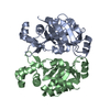

 Mass: 79.904 Da / Num. of mol.: 1 / Source method: obtained synthetically / Formula: Br
 Mass: 18.015 Da / Num. of mol.: 347 / Source method: isolated from a natural source / Formula: H2O
 Sample preparation
 / Beamline: 22-BM / Wavelength: 1.0084
  Processing