- PDB-3ii2: Structure of ORF157 from Acidianus Filamentous Virus 1 -
+
Open data
ID or keywords:
Loading...
-
Basic information
Entry
Database: PDB / ID: 3ii2
Title
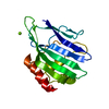
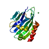
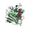
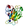
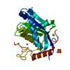
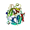
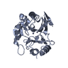
Structure of ORF157 from Acidianus Filamentous Virus 1
Components
Putative uncharacterized protein
Keywords
DNA BINDING PROTEIN / virus / archaea / nuclease
Function / homology
Protein of unknown function DUF3258 / Protein of unknown function DUF3258 / Immunoglobulin-like - #2930 / Immunoglobulin-like / Sandwich / Mainly Beta / : / NICKEL (II) ION / Uncharacterized protein ORF157
Function and homology information
Biological species
Acidianus filamentous virus 1
Method
X-RAY DIFFRACTION / SYNCHROTRON / SAD / Resolution: 2 Å
Mass: 18861.609 Da / Num. of mol.: 1 Source method: isolated from a genetically manipulated source Details: TEV cleavage site inserted between attB1 and the gene of interest Source: (gene. exp.) Acidianus filamentous virus 1 / Gene: AFV1 orf157 / Plasmid: pDEST17 / Production host: Escherichia coli (E. coli) / Strain (production host): Rosetta(DE3)pLysS / References: UniProt: Q70LE6
In the structure databanks used in Yorodumi, some data are registered as the other names, "COVID-19 virus" and "2019-nCoV". Here are the details of the virus and the list of structure data.
Jan 31, 2019. EMDB accession codes are about to change! (news from PDBe EMDB page)
EMDB accession codes are about to change! (news from PDBe EMDB page)
The allocation of 4 digits for EMDB accession codes will soon come to an end. Whilst these codes will remain in use, new EMDB accession codes will include an additional digit and will expand incrementally as the available range of codes is exhausted. The current 4-digit format prefixed with “EMD-” (i.e. EMD-XXXX) will advance to a 5-digit format (i.e. EMD-XXXXX), and so on. It is currently estimated that the 4-digit codes will be depleted around Spring 2019, at which point the 5-digit format will come into force.
The EM Navigator/Yorodumi systems omit the EMD- prefix.
Related info.:Q: What is EMD? / ID/Accession-code notation in Yorodumi/EM Navigator
Yorodumi is a browser for structure data from EMDB, PDB, SASBDB, etc.
This page is also the successor to EM Navigator detail page, and also detail information page/front-end page for Omokage search.
The word "yorodu" (or yorozu) is an old Japanese word meaning "ten thousand". "mi" (miru) is to see.
Related info.:EMDB / PDB / SASBDB / Comparison of 3 databanks / Yorodumi Search / Aug 31, 2016. New EM Navigator & Yorodumi / Yorodumi Papers / Jmol/JSmol / Function and homology information / Changes in new EM Navigator and Yorodumi
 Movie
Movie Controller


 Open data
 Basic information
 Components
 Keywords
 Function and homology information
 Acidianus filamentous virus 1
 X-RAY DIFFRACTION /
  Authors
 Citation
 Structure visualization
 Downloads & links
 Other downloads
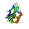


 PDBj
PDBj


 Assembly
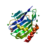


 Mass: 200.590 Da / Num. of mol.: 2 / Source method: obtained synthetically / Formula: Hg
 Mass: 35.453 Da / Num. of mol.: 3 / Source method: obtained synthetically / Formula: Cl
 Mass: 58.693 Da / Num. of mol.: 3 / Source method: obtained synthetically / Formula: Ni
 Mass: 92.094 Da / Num. of mol.: 1 / Source method: obtained synthetically / Formula: C3H8O3
 Sample preparation
 / Beamline: ID14-4 / Wavelength: 1.005 Å
 Processing