 Movie
Movie Controller


+ Open data

- Basic information
| Entry | Database: PDB / ID: 1pl3 | ||||||||||||
|---|---|---|---|---|---|---|---|---|---|---|---|---|---|
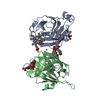
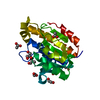
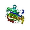
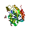
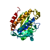
| Title | Cytochrome Domain Of Cellobiose Dehydrogenase, M65H mutant | ||||||||||||
 Components Components | Cellobiose dehydrogenase | ||||||||||||
 Keywords Keywords | OXIDOREDUCTASE / B-TYPE CYTOCHROME / MUTANT / BIS-HIS LIGATION / BETA SANDWICH / FE(II)-PROTOPORPHYRIN IX | ||||||||||||
| Function / homology |  Function and homology information Function and homology informationcellobiose dehydrogenase (acceptor) / cellobiose dehydrogenase (acceptor) activity / cellulose catabolic process / flavin adenine dinucleotide binding / extracellular region / metal ion binding Similarity search - Function | ||||||||||||
| Biological species |  Phanerochaete chrysosporium (fungus) Phanerochaete chrysosporium (fungus) | ||||||||||||
| Method |  X-RAY DIFFRACTION / SYNCHROTRON / FOURIER SYNTHESIS / Resolution: 1.9 Å X-RAY DIFFRACTION / SYNCHROTRON / FOURIER SYNTHESIS / Resolution: 1.9 Å | ||||||||||||
 Authors Authors | Rotsaert, F.A.J. / Hallberg, B.M. / de Vries, S. / Moenne-Loccoz, P. / Divne, C. / Gold, M.H. | ||||||||||||
 Citation Citation | Journal: J.Biol.Chem. / Year: 2003 Title: Biophysical and Structural Analysis of a Novel Heme b Iron Ligation in the Flavocytochrome Cellobiose Dehydrogenase. Authors: Rotsaert, F.A.J. / Hallberg, B.M. / De Vries, S. / Moenne-Loccoz, P. / Divne, C. / Renganathan, V. / Gold, M.H. | ||||||||||||
| History |
|
- Structure visualization
| Structure viewer | Molecule: MolmilJmol/JSmol |
|---|
- Downloads & links
-Download
| PDBx/mmCIF format | 1pl3.cif.gz | 97.6 KB | Display | PDBx/mmCIF format |
|---|---|---|---|---|
| PDB format | pdb1pl3.ent.gz | 73.7 KB | Display | PDB format |
| PDBx/mmJSON format | 1pl3.json.gz | Tree view | PDBx/mmJSON format | |
| Others |  Other downloads Other downloads |
-Validation report
| Arichive directory | https://data.pdbj.org/pub/pdb/validation_reports/pl/1pl3ftp://data.pdbj.org/pub/pdb/validation_reports/pl/1pl3 | HTTPS FTP |
|---|
-Related structure data
| Related structure data |  1d7cS S: Starting model for refinement |
|---|---|
| Similar structure data |


-Links
 PDBj
PDBj
- Assembly
| Deposited unit | 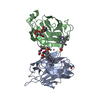
| ||||||||
|---|---|---|---|---|---|---|---|---|---|
| 1 | 
| ||||||||
| 2 | 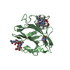
| ||||||||
| Unit cell |
|
-Components
-Protein / Sugars , 2 types, 4 molecules AB
| #1: Protein | Mass: 20019.719 Da / Num. of mol.: 2 / Fragment: CYTOCHROME TYPE B HEME DOMAIN / Mutation: M65H Source method: isolated from a genetically manipulated source Source: (gene. exp.) Phanerochaete chrysosporium (fungus) / Gene: CDH-1 AND CDH-2 / Plasmid: pUGC1 / Production host: Phanerochaete chrysosporium (fungus) / Strain (production host): OGC316-7References: UniProt: Q01738, cellobiose dehydrogenase (acceptor) #2: Polysaccharide | Source method: isolated from a genetically manipulated source |
|---|
-Non-polymers , 4 types, 345 molecules 


| #3: Chemical | ChemComp-CD /  Mass: 112.411 Da / Num. of mol.: 6 / Source method: obtained synthetically / Formula: Cd Mass: 112.411 Da / Num. of mol.: 6 / Source method: obtained synthetically / Formula: Cd#4: Chemical |  Mass: 616.487 Da / Num. of mol.: 2 / Source method: obtained synthetically / Formula: C34H32FeN4O4 Mass: 616.487 Da / Num. of mol.: 2 / Source method: obtained synthetically / Formula: C34H32FeN4O4#5: Chemical | ChemComp-1PG / |  Mass: 252.305 Da / Num. of mol.: 1 / Source method: obtained synthetically / Formula: C11H24O6 Mass: 252.305 Da / Num. of mol.: 1 / Source method: obtained synthetically / Formula: C11H24O6#6: Water | ChemComp-HOH / | Mass: 18.015 Da / Num. of mol.: 336 / Source method: isolated from a natural source / Formula: H2O |
|---|
-Details
| Has protein modification | Y |
|---|
-Experimental details
-Experiment
| Experiment | Method: X-RAY DIFFRACTION / Number of used crystals: 1 |
|---|
- Sample preparation
| Crystal | Density Matthews: 3.67 Å3/Da / Density % sol: 66.47 % | ||||||||||||||||||||||||||||||||||||||||||
|---|---|---|---|---|---|---|---|---|---|---|---|---|---|---|---|---|---|---|---|---|---|---|---|---|---|---|---|---|---|---|---|---|---|---|---|---|---|---|---|---|---|---|---|
| Crystal grow | Temperature: 293 K / Method: vapor diffusion, hanging drop / pH: 7.5 Details: PEG 4000, 2-METHYL 2,4-PENTANEDIOL, HEPES, CADMIUM CHLORIDE, pH 7.5, VAPOR DIFFUSION, HANGING DROP, temperature 293K | ||||||||||||||||||||||||||||||||||||||||||
| Crystal grow | *PLUS Method: vapor diffusion, hanging drop | ||||||||||||||||||||||||||||||||||||||||||
| Components of the solutions | *PLUS
|
-Data collection
| Diffraction | Mean temperature: 100 K |
|---|---|
| Diffraction source | Source: SYNCHROTRON / Site: ESRF  / Beamline: ID14-4 / Wavelength: 0.9763 Å / Beamline: ID14-4 / Wavelength: 0.9763 Å |
| Detector | Type: ADSC QUANTUM 4 / Detector: CCD / Date: May 4, 2002 |
| Radiation | Protocol: SINGLE WAVELENGTH / Monochromatic (M) / Laue (L): M / Scattering type: x-ray |
| Radiation wavelength | Wavelength: 0.9763 Å / Relative weight: 1 |
| Reflection | Resolution: 1.9→48 Å / Num. all: 45580 / Num. obs: 45580 / % possible obs: 99.4 % / Observed criterion σ(F): 0 / Observed criterion σ(I): 0 / Redundancy: 5.6 % / Rmerge(I) obs: 0.113 / Net I/σ(I): 5 |
| Reflection shell | Resolution: 1.9→2 Å / Redundancy: 4.9 % / Rmerge(I) obs: 0.575 / Mean I/σ(I) obs: 1.1 / % possible all: 97.5 |
| Reflection | *PLUS Num. obs: 45850 / Num. measured all: 256083 |
| Reflection shell | *PLUS % possible obs: 97.5 % |
- Processing
| Software |
| |||||||||||||||||||||||||
|---|---|---|---|---|---|---|---|---|---|---|---|---|---|---|---|---|---|---|---|---|---|---|---|---|---|---|
| Refinement | Method to determine structure: FOURIER SYNTHESIS Starting model: 1D7C Resolution: 1.9→28 Å / Cross valid method: THROUGHOUT / σ(F): 0 / Stereochemistry target values: Engh & Huber
| |||||||||||||||||||||||||
| Refinement step | Cycle: LAST / Resolution: 1.9→28 Å
| |||||||||||||||||||||||||
| Refine LS restraints |
| |||||||||||||||||||||||||
| LS refinement shell | Resolution: 1.9→1.949 Å
| |||||||||||||||||||||||||
| Refinement | *PLUS % reflection Rfree: 5 % / Rfactor Rwork: 0.173 | |||||||||||||||||||||||||
| Solvent computation | *PLUS | |||||||||||||||||||||||||
| Displacement parameters | *PLUS | |||||||||||||||||||||||||
| Refine LS restraints | *PLUS
|
