 Movie
Movie Controller

[English] 日本語
 Yorodumi
- PDB-1kdg: Crystal structure of the flavin domain of cellobiose dehydrogenase -
+ Open data

- Basic information
| Entry | Database: PDB / ID: 1kdg | ||||||
|---|---|---|---|---|---|---|---|
| Title | Crystal structure of the flavin domain of cellobiose dehydrogenase | ||||||
 Components Components | cellobiose dehydrogenase | ||||||
 Keywords Keywords | OXIDOREDUCTASE / GMC oxidoreductase / PHBH fold / alpha/beta structure / Rossmann fold / 6-hydroxylated FAD | ||||||
| Function / homology |  Function and homology information Function and homology informationcellobiose dehydrogenase (acceptor) / cellobiose dehydrogenase (acceptor) activity / cellulose catabolic process / flavin adenine dinucleotide binding / extracellular region / metal ion binding Similarity search - Function | ||||||
| Biological species |  Phanerochaete chrysosporium (fungus) Phanerochaete chrysosporium (fungus) | ||||||
| Method |  X-RAY DIFFRACTION / SYNCHROTRON / MAD / Resolution: 1.5 Å X-RAY DIFFRACTION / SYNCHROTRON / MAD / Resolution: 1.5 Å | ||||||
 Authors Authors | Hallberg, B.M. / Henriksson, G. / Pettersson, G. / Divne, C. | ||||||
 Citation Citation | Journal: J.Mol.Biol. / Year: 2002 Title: Crystal Structure of the Flavoprotein Domain of the Extracellular Flavocytochrome Cellobiose Dehydrogenase Authors: Hallberg, B.M. / Henriksson, G. / Pettersson, G. / Divne, C. | ||||||
| History |
|
- Structure visualization
| Structure viewer | Molecule: MolmilJmol/JSmol |
|---|
- Downloads & links
-Download
| PDBx/mmCIF format | 1kdg.cif.gz | 465.9 KB | Display | PDBx/mmCIF format |
|---|---|---|---|---|
| PDB format | pdb1kdg.ent.gz | 377.4 KB | Display | PDB format |
| PDBx/mmJSON format | 1kdg.json.gz | Tree view | PDBx/mmJSON format | |
| Others |  Other downloads Other downloads |
-Validation report
| Arichive directory | https://data.pdbj.org/pub/pdb/validation_reports/kd/1kdgftp://data.pdbj.org/pub/pdb/validation_reports/kd/1kdg | HTTPS FTP |
|---|
-Related structure data
| Related structure data | |
|---|---|
| Similar structure data |


-Links
 PDBj
PDBj


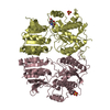
- Assembly
| Deposited unit | 
| ||||||||||
|---|---|---|---|---|---|---|---|---|---|---|---|
| 1 |
| ||||||||||
| 2 | 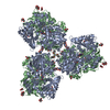
| ||||||||||
| Unit cell |
| ||||||||||
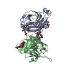
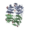
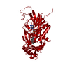
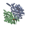
| Details | THE BIOLOGICAL ASSEMBLY IS A MONOMERIC FLAVOCYTOCHROME CONSISTING OF A B-TYPE CYTOCHROME DOMAIN LINKED TO A FLAVODEHYDROGENASE DOMAIN |
-Components
-Protein , 1 types, 2 molecules AB
| #1: Protein | Mass: 58000.473 Da / Num. of mol.: 2 / Fragment: C-terminal flavoprotein domain / Source method: isolated from a natural source / Details: secreted / Source: (natural) Phanerochaete chrysosporium (fungus) / Strain: K3References: UniProt: Q01738, cellobiose dehydrogenase (acceptor) |
|---|
-Sugars , 2 types, 9 molecules 

| #2: Sugar | ChemComp-NAG /  Type: D-saccharide, beta linking / Mass: 221.208 Da / Num. of mol.: 5 Type: D-saccharide, beta linking / Mass: 221.208 Da / Num. of mol.: 5Source method: isolated from a genetically manipulated source Formula: C8H15NO6 #3: Sugar | ChemComp-MAN /  Type: D-saccharide, alpha linking / Mass: 180.156 Da / Num. of mol.: 4 Type: D-saccharide, alpha linking / Mass: 180.156 Da / Num. of mol.: 4Source method: isolated from a genetically manipulated source Formula: C6H12O6 |
|---|
-Non-polymers , 5 types, 1133 molecules 
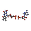


| #4: Chemical | ChemComp-HG /  Mass: 200.590 Da / Num. of mol.: 9 / Source method: obtained synthetically / Formula: Hg Mass: 200.590 Da / Num. of mol.: 9 / Source method: obtained synthetically / Formula: Hg#5: Chemical |  Mass: 801.549 Da / Num. of mol.: 2 / Source method: obtained synthetically / Formula: C27H33N9O16P2 Mass: 801.549 Da / Num. of mol.: 2 / Source method: obtained synthetically / Formula: C27H33N9O16P2#6: Chemical | ChemComp-EMT / |  Mass: 382.830 Da / Num. of mol.: 1 / Source method: obtained synthetically / Formula: C9H10HgO2S Mass: 382.830 Da / Num. of mol.: 1 / Source method: obtained synthetically / Formula: C9H10HgO2S#7: Chemical | ChemComp-UNX / |  Num. of mol.: 1 / Source method: obtained synthetically Num. of mol.: 1 / Source method: obtained synthetically#8: Water | ChemComp-HOH / | Mass: 18.015 Da / Num. of mol.: 1120 / Source method: isolated from a natural source / Formula: H2O |
|---|
-Details
| Has protein modification | Y |
|---|
-Experimental details
-Experiment
| Experiment | Method: X-RAY DIFFRACTION / Number of used crystals: 1 |
|---|
- Sample preparation
| Crystal | Density Matthews: 2.3 Å3/Da / Density % sol: 46.54 % | ||||||||||||||||||||||||||||||
|---|---|---|---|---|---|---|---|---|---|---|---|---|---|---|---|---|---|---|---|---|---|---|---|---|---|---|---|---|---|---|---|
| Crystal grow | Temperature: 288 K / Method: vapor diffusion, hanging drop / pH: 6.5 Details: AMMONIUM SULFATE, DIOXANE, MES, MERTHIOLATE, pH 6.5, VAPOR DIFFUSION, HANGING DROP at 288K | ||||||||||||||||||||||||||||||
| Crystal grow | *PLUS Temperature: 15 ℃ | ||||||||||||||||||||||||||||||
| Components of the solutions | *PLUS
|
-Data collection
| Diffraction | Mean temperature: 100 K |
|---|---|
| Diffraction source | Source: SYNCHROTRON / Site: MAX II  / Beamline: I711 / Wavelength: 0.996 Å / Beamline: I711 / Wavelength: 0.996 Å |
| Detector | Type: MARRESEARCH / Detector: IMAGE PLATE / Date: Jan 17, 1999 |
| Radiation | Monochromator: Si 111 CHANNEL / Protocol: SINGLE WAVELENGTH / Monochromatic (M) / Laue (L): M / Scattering type: x-ray |
| Radiation wavelength | Wavelength: 0.996 Å / Relative weight: 1 |
| Reflection | Resolution: 1.5→25 Å / Num. all: 165042 / Num. obs: 165042 / % possible obs: 99.6 % / Observed criterion σ(F): 0 / Observed criterion σ(I): 0 / Redundancy: 2.3 % / Rmerge(I) obs: 0.08 / Net I/σ(I): 4.7 |
| Reflection shell | Resolution: 1.5→1.58 Å / Rmerge(I) obs: 0.451 / Mean I/σ(I) obs: 1.2 / % possible all: 99.2 |
| Reflection | *PLUS Lowest resolution: 25 Å / Num. measured all: 385729 / Rmerge(I) obs: 0.08 |
| Reflection shell | *PLUS % possible obs: 99.2 % |
- Processing
| Software |
| |||||||||||||||||||||||||
|---|---|---|---|---|---|---|---|---|---|---|---|---|---|---|---|---|---|---|---|---|---|---|---|---|---|---|
| Refinement | Method to determine structure: MAD / Resolution: 1.5→23 Å / Isotropic thermal model: anisotropic / Cross valid method: THROUGHOUT / σ(F): 0 / Stereochemistry target values: Engh & Huber
| |||||||||||||||||||||||||
| Refinement step | Cycle: LAST / Resolution: 1.5→23 Å
| |||||||||||||||||||||||||
| Refine LS restraints |
| |||||||||||||||||||||||||
| LS refinement shell | Resolution: 1.5→1.539 Å / Rfactor Rfree error: 0.036
| |||||||||||||||||||||||||
| Refinement | *PLUS Lowest resolution: 23 Å / Num. reflection obs: 162564 / Rfactor Rwork: 0.134 | |||||||||||||||||||||||||
| Solvent computation | *PLUS | |||||||||||||||||||||||||
| Displacement parameters | *PLUS |
