 Movie
Movie Controller


+ Open data

- Basic information
| Entry | Database: PDB / ID: 3ii3 | ||||||
|---|---|---|---|---|---|---|---|
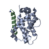
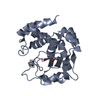
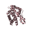
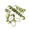
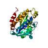
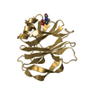
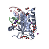
| Title | Structure of ORF157 from Acidianus filamentous Virus 1 | ||||||
 Components Components | Putative uncharacterized protein | ||||||
 Keywords Keywords | DNA BINDING PROTEIN / virus / archaea / nuclease | ||||||
| Function / homology | Protein of unknown function DUF3258 / Protein of unknown function DUF3258 / Immunoglobulin-like - #2930 / Immunoglobulin-like / Sandwich / Mainly Beta / NICKEL (II) ION / Uncharacterized protein ORF157 Function and homology information Function and homology information | ||||||
| Biological species |  Acidianus filamentous virus 1 Acidianus filamentous virus 1 | ||||||
| Method |  X-RAY DIFFRACTION / SYNCHROTRON / MOLECULAR REPLACEMENT / Resolution: 2.7 Å X-RAY DIFFRACTION / SYNCHROTRON / MOLECULAR REPLACEMENT / Resolution: 2.7 Å | ||||||
 Authors Authors | Goulet, A. / Redder, P. / Pina, M. / Prangishvili, D. / van Tilbeurgh, H. / Cambillau, C. / Campanacci, V. | ||||||
 Citation Citation | Journal: J.Virol. / Year: 2010 Title: ORF157 from the archaeal virus Acidianus filamentous virus 1 defines a new class of nuclease Authors: Goulet, A. / Pina, M. / Redder, P. / Prangishvili, D. / Vera, L. / Lichiere, J. / Leulliot, N. / van Tilbeurgh, H. / Ortiz-Lombardia, M. / Campanacci, V. / Cambillau, C. | ||||||
| History |
|
- Structure visualization
| Structure viewer | Molecule: MolmilJmol/JSmol |
|---|
- Downloads & links
-Download
| PDBx/mmCIF format | 3ii3.cif.gz | 44.9 KB | Display | PDBx/mmCIF format |
|---|---|---|---|---|
| PDB format | pdb3ii3.ent.gz | 31 KB | Display | PDB format |
| PDBx/mmJSON format | 3ii3.json.gz | Tree view | PDBx/mmJSON format | |
| Others |  Other downloads Other downloads |
-Validation report
| Arichive directory | https://data.pdbj.org/pub/pdb/validation_reports/ii/3ii3ftp://data.pdbj.org/pub/pdb/validation_reports/ii/3ii3 | HTTPS FTP |
|---|
-Related structure data
| Related structure data | 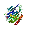 3ii2SC 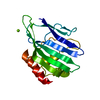 3ildC 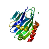 3ileC S: Starting model for refinement C: citing same article ( |
|---|---|
| Similar structure data |


-Links
 PDBj
PDBj- Assembly
| Deposited unit | 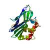
| |||||||||
|---|---|---|---|---|---|---|---|---|---|---|
| 1 |
| |||||||||
| Unit cell |
| |||||||||
| Components on special symmetry positions |
|
-Components
| #1: Protein | Mass: 18861.609 Da / Num. of mol.: 1 Source method: isolated from a genetically manipulated source Details: TEV cleavage site inserted between attB1 and the gene of interest Source: (gene. exp.) Acidianus filamentous virus 1 / Gene: AFV1 orf157 / Plasmid: pDEST17 / Production host:  | ||
|---|---|---|---|
| #2: Chemical |   Mass: 58.693 Da / Num. of mol.: 2 / Source method: obtained synthetically / Formula: Ni Mass: 58.693 Da / Num. of mol.: 2 / Source method: obtained synthetically / Formula: Ni#3: Chemical | ChemComp-GOL / |   Mass: 92.094 Da / Num. of mol.: 1 / Source method: obtained synthetically / Formula: C3H8O3 Mass: 92.094 Da / Num. of mol.: 1 / Source method: obtained synthetically / Formula: C3H8O3 |
-Experimental details
-Experiment
| Experiment | Method: X-RAY DIFFRACTION / Number of used crystals: 1 |
|---|
- Sample preparation
| Crystal | Density Matthews: 2.81 Å3/Da / Density % sol: 56.16 % |
|---|---|
| Crystal grow | Temperature: 293 K / Method: vapor diffusion, sitting drop / pH: 8 Details: 0.9M LiSO4, 0.01M NiCl2, 0.1M Tris, pH 8, VAPOR DIFFUSION, SITTING DROP, temperature 293K |
-Data collection
| Diffraction | Mean temperature: 100 K | |||||||||||||||
|---|---|---|---|---|---|---|---|---|---|---|---|---|---|---|---|---|
| Diffraction source | Source: SYNCHROTRON / Site: ESRF  / Beamline: ID14-3 / Wavelength: 0.93 Å / Beamline: ID14-3 / Wavelength: 0.93 Å | |||||||||||||||
| Detector | Type: MAR CCD 165 mm / Detector: CCD / Date: Feb 9, 2007 | |||||||||||||||
| Radiation | Protocol: SINGLE WAVELENGTH / Monochromatic (M) / Laue (L): M / Scattering type: x-ray | |||||||||||||||
| Radiation wavelength | Wavelength: 0.93 Å / Relative weight: 1 | |||||||||||||||
| Reflection twin |
| |||||||||||||||
| Reflection | Resolution: 2.7→40 Å / Num. obs: 6151 / % possible obs: 100 % / Redundancy: 5.2 % / Biso Wilson estimate: 78.4 Å2 / Rmerge(I) obs: 0.084 / Net I/σ(I): 10.3 | |||||||||||||||
| Reflection shell | Resolution: 2.7→2.85 Å / Redundancy: 5.3 % / Rmerge(I) obs: 0.46 / Mean I/σ(I) obs: 3.1 / Num. unique all: 873 / % possible all: 100 |
- Processing
| Software |
| |||||||||||||||||||||||||||||||||||||||||||||||||||||||||||||||||
|---|---|---|---|---|---|---|---|---|---|---|---|---|---|---|---|---|---|---|---|---|---|---|---|---|---|---|---|---|---|---|---|---|---|---|---|---|---|---|---|---|---|---|---|---|---|---|---|---|---|---|---|---|---|---|---|---|---|---|---|---|---|---|---|---|---|---|
| Refinement | Method to determine structure: MOLECULAR REPLACEMENT Starting model: PDB entry 3II2 Resolution: 2.7→25 Å / Cor.coef. Fo:Fc: 0.907 / Cor.coef. Fo:Fc free: 0.916 / SU B: 41.278 / SU ML: 0.367 / TLS residual ADP flag: LIKELY RESIDUAL / Cross valid method: THROUGHOUT / ESU R: 0.242 / ESU R Free: 0.078 / Stereochemistry target values: MAXIMUM LIKELIHOOD
| |||||||||||||||||||||||||||||||||||||||||||||||||||||||||||||||||
| Solvent computation | Ion probe radii: 0.8 Å / Shrinkage radii: 0.8 Å / VDW probe radii: 1.4 Å / Solvent model: MASK | |||||||||||||||||||||||||||||||||||||||||||||||||||||||||||||||||
| Displacement parameters | Biso mean: 30.744 Å2
| |||||||||||||||||||||||||||||||||||||||||||||||||||||||||||||||||
| Refinement step | Cycle: LAST / Resolution: 2.7→25 Å
| |||||||||||||||||||||||||||||||||||||||||||||||||||||||||||||||||
| Refine LS restraints |
| |||||||||||||||||||||||||||||||||||||||||||||||||||||||||||||||||
| LS refinement shell | Resolution: 2.699→2.768 Å / Total num. of bins used: 20
| |||||||||||||||||||||||||||||||||||||||||||||||||||||||||||||||||
| Refinement TLS params. | Method: refined / Origin x: 27.1227 Å / Origin y: 16.2577 Å / Origin z: 5.4136 Å
|
