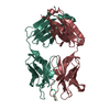
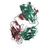
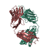
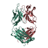
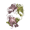
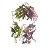
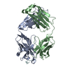
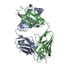
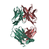
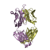
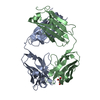
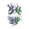
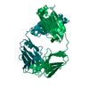
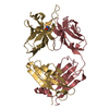
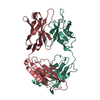
Entry Database : PDB / ID : 3iflTitle X-ray structure of amyloid beta peptide:antibody (Abeta1-7:12A11) complex 12A11 FAB antibody heavy chain 12A11 FAB antibody light chain Amyloid beta A4 protein Keywords / / Function / homology Function Domain/homology Component
/ / / / / / / / / / / / / / / / / / / / / / / / / / / / / / / / / / / / / / / / / / / / / / / / / / / / / / / / / / / / / / / / / / / / / / / / / / / / / / / / / / / / / / / / / / / / / / / / / / / / / / / / / / / / / / / / / / / / / / / / / / / / / / / / / / / / / / / / / / / / / / / / / / / / / / / Biological species Mus musculus (house mouse)Homo sapiens (human)Method / / / Resolution : 1.5 Å Authors Weis, W.I. / Feinberg, H. / Basi, G.S. / Schenk, D. Journal : J.Biol.Chem. / Year : 2010Title : Structural correlates of antibodies associated with acute reversal of amyloid beta-related behavioral deficits in a mouse model of Alzheimer disease.Authors: Basi, G.S. / Feinberg, H. / Oshidari, F. / Anderson, J. / Barbour, R. / Baker, J. / Comery, T.A. / Diep, L. / Gill, D. / Johnson-Wood, K. / Goel, A. / Grantcharova, K. / Lee, M. / Li, J. / ... Authors : Basi, G.S. / Feinberg, H. / Oshidari, F. / Anderson, J. / Barbour, R. / Baker, J. / Comery, T.A. / Diep, L. / Gill, D. / Johnson-Wood, K. / Goel, A. / Grantcharova, K. / Lee, M. / Li, J. / Partridge, A. / Griswold-Prenner, I. / Piot, N. / Walker, D. / Widom, A. / Pangalos, M.N. / Seubert, P. / Jacobsen, J.S. / Schenk, D. / Weis, W.I. History Deposition Jul 24, 2009 Deposition site / Processing site Revision 1.0 Nov 17, 2009 Provider / Type Revision 1.1 Jul 13, 2011 Group Revision 1.2 Jun 19, 2013 Group Revision 1.3 Nov 20, 2024 Group / Database references / Structure summaryCategory chem_comp_atom / chem_comp_bond ... chem_comp_atom / chem_comp_bond / database_2 / pdbx_entry_details / pdbx_modification_feature Item / _database_2.pdbx_database_accession
Show all Show less
 Movie
Movie Controller

 Yorodumi
 Open data
 Basic information
 Components
 Keywords
 Function and homology information
 Homo sapiens (human)
 X-RAY DIFFRACTION /
  Authors
 Citation
 Structure visualization
 Downloads & links
 Other downloads


 PDBj
PDBj


 Assembly
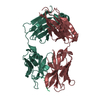
 Cricetulus griseus (Chinese hamster) / Tissue (production host): OVARY
 Mass: 18.015 Da / Num. of mol.: 582 / Source method: isolated from a natural source / Formula: H2O
 Sample preparation
 / Beamline: BL11-1 / Wavelength: 0.97946 Å
 Processing