 Movie
Movie Controller


+ Open data

- Basic information
| Entry | Database: PDB / ID: 3hz2 | ||||||
|---|---|---|---|---|---|---|---|
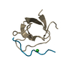
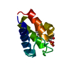
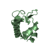
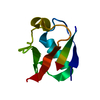
| Title | Crystal structure of a betagamma-crystallin from an Archaea | ||||||
 Components Components | Beta/gama crystallin family protein | ||||||
 Keywords Keywords | METAL BINDING PROTEIN / calcium-bound betagamma-crystallin | ||||||
| Function / homology |  Function and homology information Function and homology information | ||||||
| Biological species |  Methanosarcina acetivorans (archaea) Methanosarcina acetivorans (archaea) | ||||||
| Method |  X-RAY DIFFRACTION / MOLECULAR REPLACEMENT / molecular replacement / Resolution: 1.86 Å X-RAY DIFFRACTION / MOLECULAR REPLACEMENT / molecular replacement / Resolution: 1.86 Å | ||||||
 Authors Authors | Aravind, P. / Sankaranarayanan, R. | ||||||
 Citation Citation | Journal: Biochemistry / Year: 2009 Title: The betagamma-crystallin superfamily contains a universal motif for binding calcium Authors: Aravind, P. / Mishra, A. / Suman, S.K. / Jobby, M.K. / Sankaranarayanan, R. / Sharma, Y. | ||||||
| History |
|
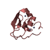
- Structure visualization
| Structure viewer | Molecule: MolmilJmol/JSmol |
|---|
- Downloads & links
-Download
| PDBx/mmCIF format | 3hz2.cif.gz | 89.7 KB | Display | PDBx/mmCIF format |
|---|---|---|---|---|
| PDB format | pdb3hz2.ent.gz | 66.6 KB | Display | PDB format |
| PDBx/mmJSON format | 3hz2.json.gz | Tree view | PDBx/mmJSON format | |
| Others |  Other downloads Other downloads |
-Validation report
| Summary document | 3hz2_validation.pdf.gz | 444.2 KB | Display | wwPDB validaton report |
|---|---|---|---|---|
| Full document | 3hz2_full_validation.pdf.gz | 445.9 KB | Display | |
| Data in XML | 3hz2_validation.xml.gz | 19.5 KB | Display | |
| Data in CIF | 3hz2_validation.cif.gz | 28.5 KB | Display | |
| Arichive directory | https://data.pdbj.org/pub/pdb/validation_reports/hz/3hz2ftp://data.pdbj.org/pub/pdb/validation_reports/hz/3hz2 | HTTPS FTP |
-Related structure data
| Related structure data |  3hzbC  3i9hC  3iajC  2bv2S S: Starting model for refinement C: citing same article ( |
|---|---|
| Similar structure data |


-Links
 PDBj
PDBj
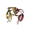
- Assembly
| Deposited unit | 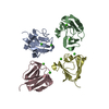
| ||||||||
|---|---|---|---|---|---|---|---|---|---|
| 1 | 
| ||||||||
| 2 | 
| ||||||||
| 3 | 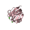
| ||||||||
| 4 | 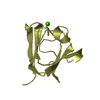
| ||||||||
| Unit cell |
|
-Components
| #1: Protein | Mass: 9252.976 Da / Num. of mol.: 4 / Fragment: UNP residues 37-120 Source method: isolated from a genetically manipulated source Source: (gene. exp.) Methanosarcina acetivorans (archaea) / Strain: C2A / Gene: 1474415 / Plasmid: pET21a / Production host:  #2: Chemical | ChemComp-CA /   Mass: 40.078 Da / Num. of mol.: 8 / Source method: obtained synthetically / Formula: Ca Mass: 40.078 Da / Num. of mol.: 8 / Source method: obtained synthetically / Formula: Ca#3: Water | ChemComp-HOH / |  Mass: 18.015 Da / Num. of mol.: 450 / Source method: isolated from a natural source / Formula: H2O Mass: 18.015 Da / Num. of mol.: 450 / Source method: isolated from a natural source / Formula: H2O |
|---|
-Experimental details
-Experiment
| Experiment | Method: X-RAY DIFFRACTION / Number of used crystals: 1 |
|---|
- Sample preparation
| Crystal | Density Matthews: 2.15 Å3/Da / Density % sol: 42.92 % / Mosaicity: 0.594 ° |
|---|---|
| Crystal grow | Temperature: 298 K / Method: vapor diffusion, sitting drop / pH: 6.5 Details: 12% PEG 20000, 0.1M NaMES, pH6.5, temperature 298K, VAPOR DIFFUSION, SITTING DROP |
-Data collection
| Diffraction | Mean temperature: 100 K | |||||||||||||||||||||||||||||||||||||||||||||||||||||||||||||||||||||||||||||
|---|---|---|---|---|---|---|---|---|---|---|---|---|---|---|---|---|---|---|---|---|---|---|---|---|---|---|---|---|---|---|---|---|---|---|---|---|---|---|---|---|---|---|---|---|---|---|---|---|---|---|---|---|---|---|---|---|---|---|---|---|---|---|---|---|---|---|---|---|---|---|---|---|---|---|---|---|---|---|
| Diffraction source | Source: ROTATING ANODE / Type: RIGAKU RUH3R / Wavelength: 1.54 Å | |||||||||||||||||||||||||||||||||||||||||||||||||||||||||||||||||||||||||||||
| Detector | Type: MAR scanner 345 mm plate / Detector: IMAGE PLATE / Date: Jan 29, 2008 | |||||||||||||||||||||||||||||||||||||||||||||||||||||||||||||||||||||||||||||
| Radiation | Protocol: SINGLE WAVELENGTH / Monochromatic (M) / Laue (L): M / Scattering type: x-ray | |||||||||||||||||||||||||||||||||||||||||||||||||||||||||||||||||||||||||||||
| Radiation wavelength | Wavelength: 1.54 Å / Relative weight: 1 | |||||||||||||||||||||||||||||||||||||||||||||||||||||||||||||||||||||||||||||
| Reflection | Resolution: 1.86→25 Å / Num. obs: 24461 / % possible obs: 94.1 % / Redundancy: 2.6 % / Rmerge(I) obs: 0.037 / Χ2: 1.176 / Net I/σ(I): 28.857 | |||||||||||||||||||||||||||||||||||||||||||||||||||||||||||||||||||||||||||||
| Reflection shell |
|
-Phasing
| Phasing | Method: molecular replacement |
|---|
- Processing
| Software |
| ||||||||||||||||||||||||
|---|---|---|---|---|---|---|---|---|---|---|---|---|---|---|---|---|---|---|---|---|---|---|---|---|---|
| Refinement | Method to determine structure: MOLECULAR REPLACEMENT Starting model: PDB ENTRY 2BV2 Resolution: 1.86→25 Å / Occupancy max: 1 / Occupancy min: 1 / Cross valid method: THROUGHOUT / σ(F): 0
| ||||||||||||||||||||||||
| Solvent computation | Bsol: 42.207 Å2 | ||||||||||||||||||||||||
| Displacement parameters | Biso max: 55.4 Å2 / Biso mean: 15.638 Å2 / Biso min: 5.04 Å2
| ||||||||||||||||||||||||
| Refinement step | Cycle: LAST / Resolution: 1.86→25 Å
| ||||||||||||||||||||||||
| Refine LS restraints |
| ||||||||||||||||||||||||
| Xplor file |
|
