 Movie
Movie Controller

[English] 日本語
 Yorodumi
- PDB-3hv8: Crystal structure of FimX EAL domain from Pseudomonas aeruginosa ... -
+ Open data

- Basic information
| Entry | Database: PDB / ID: 3hv8 | ||||||
|---|---|---|---|---|---|---|---|
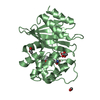
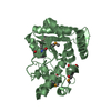
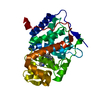
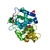
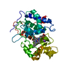
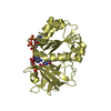
| Title | Crystal structure of FimX EAL domain from Pseudomonas aeruginosa bound to c-di-GMP | ||||||
 Components Components | Protein FimX | ||||||
 Keywords Keywords | HYDROLASE / EAL phosphodiesterase / biofilm / c-di-GMP | ||||||
| Function / homology |  Function and homology information Function and homology informationregulation of single-species biofilm formation / cyclic-guanylate-specific phosphodiesterase activity / phosphorelay signal transduction system / nucleotide binding / regulation of DNA-templated transcription / metal ion binding / identical protein binding / plasma membrane Similarity search - Function | ||||||
| Biological species |  Pseudomonas aeruginosa PAO1 (bacteria) Pseudomonas aeruginosa PAO1 (bacteria) | ||||||
| Method |  X-RAY DIFFRACTION / SYNCHROTRON / MOLECULAR REPLACEMENT / Resolution: 1.445 Å X-RAY DIFFRACTION / SYNCHROTRON / MOLECULAR REPLACEMENT / Resolution: 1.445 Å | ||||||
 Authors Authors | Navarro, M.V.A.S. / De, N. / Bae, N. / Sondermann, H. | ||||||
 Citation Citation | Journal: Structure / Year: 2009 Title: Structural analysis of the GGDEF-EAL domain-containing c-di-GMP receptor FimX. Authors: Navarro, M.V. / De, N. / Bae, N. / Wang, Q. / Sondermann, H. | ||||||
| History |
|
- Structure visualization
| Structure viewer | Molecule: MolmilJmol/JSmol |
|---|
- Downloads & links
-Download
| PDBx/mmCIF format | 3hv8.cif.gz | 72.3 KB | Display | PDBx/mmCIF format |
|---|---|---|---|---|
| PDB format | pdb3hv8.ent.gz | 52.3 KB | Display | PDB format |
| PDBx/mmJSON format | 3hv8.json.gz | Tree view | PDBx/mmJSON format | |
| Others |  Other downloads Other downloads |
-Validation report
| Arichive directory | https://data.pdbj.org/pub/pdb/validation_reports/hv/3hv8ftp://data.pdbj.org/pub/pdb/validation_reports/hv/3hv8 | HTTPS FTP |
|---|
-Related structure data
| Related structure data |  3hv9C  3hvaC  4j40C  3hvb C: citing same article ( S: Starting model for refinement |
|---|---|
| Similar structure data |


-Links
 PDBj
PDBj


- Assembly
| Deposited unit | 
| ||||||||
|---|---|---|---|---|---|---|---|---|---|
| 1 |
| ||||||||
| Unit cell |
|
-Components
| #1: Protein | Mass: 29321.281 Da / Num. of mol.: 1 / Fragment: EAL domain: UNP residues 429-691 Source method: isolated from a genetically manipulated source Source: (gene. exp.) Pseudomonas aeruginosa PAO1 (bacteria) / Strain: PAO1 / 1C / PRS 101 / LMG 12228 / Gene: fimX, PA4959 / Plasmid: ppSUMO / Production host: References: UniProt: Q9HUK6, cyclic-guanylate-specific phosphodiesterase |
|---|---|
| #2: Chemical | ChemComp-C2E /   Mass: 690.411 Da / Num. of mol.: 1 / Source method: obtained synthetically / Formula: C20H24N10O14P2 Mass: 690.411 Da / Num. of mol.: 1 / Source method: obtained synthetically / Formula: C20H24N10O14P2 |
| #3: Water | ChemComp-HOH /  Mass: 18.015 Da / Num. of mol.: 318 / Source method: isolated from a natural source / Formula: H2O Mass: 18.015 Da / Num. of mol.: 318 / Source method: isolated from a natural source / Formula: H2O |
-Experimental details
-Experiment
| Experiment | Method: X-RAY DIFFRACTION / Number of used crystals: 1 |
|---|
- Sample preparation
| Crystal | Density Matthews: 2.61 Å3/Da / Density % sol: 52.9 % |
|---|---|
| Crystal grow | Temperature: 298 K / Method: vapor diffusion, hanging drop / pH: 7.1 Details: Ammonium sulfate, PEG 8000, pH 7.1, VAPOR DIFFUSION, HANGING DROP, temperature 298.0K |
-Data collection
| Diffraction | Mean temperature: 100 K |
|---|---|
| Diffraction source | Source: SYNCHROTRON / Site: CHESS  / Beamline: F1 / Wavelength: 0.918 Å / Beamline: F1 / Wavelength: 0.918 Å |
| Detector | Type: ADSC QUANTUM 210r / Detector: CCD / Date: Nov 2, 2008 |
| Radiation | Monochromator: Si(111) Channel / Protocol: SINGLE WAVELENGTH / Monochromatic (M) / Laue (L): M / Scattering type: x-ray |
| Radiation wavelength | Wavelength: 0.918 Å / Relative weight: 1 |
| Reflection | Resolution: 1.445→42.1 Å / Num. all: 54448 / Num. obs: 54441 / % possible obs: 98.4 % / Observed criterion σ(F): 1 / Observed criterion σ(I): 1 / Redundancy: 12.8 % / Rmerge(I) obs: 0.053 / Net I/σ(I): 25.6 |
| Reflection shell | Resolution: 1.445→1.56 Å / Redundancy: 7.8 % / Rmerge(I) obs: 0.619 / Mean I/σ(I) obs: 3.5 / Num. unique all: 8121 / % possible all: 92.3 |
- Processing
| Software |
| |||||||||||||||||||||||||||||||||||||||||||||||||||||||||||||||||||||||||||||||||||||||||||||||||||||||||||||||||||||||||||||||||||||||||||||||||||
|---|---|---|---|---|---|---|---|---|---|---|---|---|---|---|---|---|---|---|---|---|---|---|---|---|---|---|---|---|---|---|---|---|---|---|---|---|---|---|---|---|---|---|---|---|---|---|---|---|---|---|---|---|---|---|---|---|---|---|---|---|---|---|---|---|---|---|---|---|---|---|---|---|---|---|---|---|---|---|---|---|---|---|---|---|---|---|---|---|---|---|---|---|---|---|---|---|---|---|---|---|---|---|---|---|---|---|---|---|---|---|---|---|---|---|---|---|---|---|---|---|---|---|---|---|---|---|---|---|---|---|---|---|---|---|---|---|---|---|---|---|---|---|---|---|---|---|---|---|
| Refinement | Method to determine structure: MOLECULAR REPLACEMENT Starting model: PDB entry 3HVB 3hvb Resolution: 1.445→26.54 Å / SU ML: 0.89 / σ(F): 1.99 / Stereochemistry target values: ML
| |||||||||||||||||||||||||||||||||||||||||||||||||||||||||||||||||||||||||||||||||||||||||||||||||||||||||||||||||||||||||||||||||||||||||||||||||||
| Solvent computation | Shrinkage radii: 0.9 Å / VDW probe radii: 1.11 Å / Solvent model: FLAT BULK SOLVENT MODEL / Bsol: 73.618 Å2 / ksol: 0.389 e/Å3 | |||||||||||||||||||||||||||||||||||||||||||||||||||||||||||||||||||||||||||||||||||||||||||||||||||||||||||||||||||||||||||||||||||||||||||||||||||
| Refinement step | Cycle: LAST / Resolution: 1.445→26.54 Å
| |||||||||||||||||||||||||||||||||||||||||||||||||||||||||||||||||||||||||||||||||||||||||||||||||||||||||||||||||||||||||||||||||||||||||||||||||||
| Refine LS restraints |
| |||||||||||||||||||||||||||||||||||||||||||||||||||||||||||||||||||||||||||||||||||||||||||||||||||||||||||||||||||||||||||||||||||||||||||||||||||
| LS refinement shell |
|
