 Movie
Movie Controller

[English] 日本語
 Yorodumi
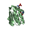
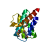
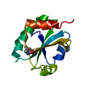
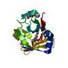
- PDB-3hrl: Crystal structure of a putative endonuclease-like protein (ngo005... -
+ Open data

- Basic information
| Entry | Database: PDB / ID: 3hrl | ||||||
|---|---|---|---|---|---|---|---|
| Title | Crystal structure of a putative endonuclease-like protein (ngo0050) from neisseria gonorrhoeae | ||||||
 Components Components | Endonuclease-Like Protein | ||||||
 Keywords Keywords | structural genomics / unknown function / ENDONUCLEASE / NEISSERIA GONORRHOEAE / PSI-2 / Protein Structure Initiative / Midwest Center for Structural Genomics / MCSG | ||||||
| Function / homology | : / Domain of unknown function DUF559 / Protein of unknown function (DUF559) / Restriction endonuclease type II-like / DNA (Cytosine-5-)-methyltransferase Function and homology information Function and homology information | ||||||
| Biological species |  Neisseria gonorrhoeae FA 1090 (bacteria) Neisseria gonorrhoeae FA 1090 (bacteria) | ||||||
| Method |  X-RAY DIFFRACTION / SYNCHROTRON / SAD / Resolution: 1.95 Å X-RAY DIFFRACTION / SYNCHROTRON / SAD / Resolution: 1.95 Å | ||||||
 Authors Authors | Filippova, E.V. / Minasov, G. / Shuvalova, L. / Kiryukhina, O. / Cobb, G. / Joachimiak, A. / Anderson, W.F. / Midwest Center for Structural Genomics (MCSG) | ||||||
 Citation Citation | Journal: To be Published Title: Crystal Structure of a Putative Endonuclease-Like Protein (Ngo0050) from Neisseria Gonorrhoeae Authors: Filippova, E.V. / Minasov, G. / Shuvalova, L. / Kiryukhina, O. / Cobb, G. / Joachimiak, A. / Anderson, W.F. | ||||||
| History |
|
- Structure visualization
| Structure viewer | Molecule: MolmilJmol/JSmol |
|---|
- Downloads & links
-Download
| PDBx/mmCIF format | 3hrl.cif.gz | 35.2 KB | Display | PDBx/mmCIF format |
|---|---|---|---|---|
| PDB format | pdb3hrl.ent.gz | 22.8 KB | Display | PDB format |
| PDBx/mmJSON format | 3hrl.json.gz | Tree view | PDBx/mmJSON format | |
| Others |  Other downloads Other downloads |
-Validation report
| Summary document | 3hrl_validation.pdf.gz | 408.5 KB | Display | wwPDB validaton report |
|---|---|---|---|---|
| Full document | 3hrl_full_validation.pdf.gz | 408.5 KB | Display | |
| Data in XML | 3hrl_validation.xml.gz | 7.8 KB | Display | |
| Data in CIF | 3hrl_validation.cif.gz | 9.9 KB | Display | |
| Arichive directory | https://data.pdbj.org/pub/pdb/validation_reports/hr/3hrlftp://data.pdbj.org/pub/pdb/validation_reports/hr/3hrl | HTTPS FTP |
-Related structure data
| Similar structure data | |
|---|---|
| Other databases |


-Links
 PDBj
PDBj


- Assembly
| Deposited unit | 
| ||||||||
|---|---|---|---|---|---|---|---|---|---|
| 1 |
| ||||||||
| Unit cell |
|
-Components
| #1: Protein | Mass: 12215.399 Da / Num. of mol.: 1 Source method: isolated from a genetically manipulated source Source: (gene. exp.) Neisseria gonorrhoeae FA 1090 (bacteria)Gene: NGO0050 / Plasmid: pMCSG7 / Production host: |
|---|---|
| #2: Chemical | ChemComp-CL /   Mass: 35.453 Da / Num. of mol.: 1 / Source method: obtained synthetically / Formula: Cl Mass: 35.453 Da / Num. of mol.: 1 / Source method: obtained synthetically / Formula: Cl |
| #3: Water | ChemComp-HOH /  Mass: 18.015 Da / Num. of mol.: 105 / Source method: isolated from a natural source / Formula: H2O Mass: 18.015 Da / Num. of mol.: 105 / Source method: isolated from a natural source / Formula: H2O |
| Has protein modification | Y |
-Experimental details
-Experiment
| Experiment | Method: X-RAY DIFFRACTION / Number of used crystals: 1 |
|---|
- Sample preparation
| Crystal | Density Matthews: 3.39 Å3/Da / Density % sol: 63.67 % |
|---|---|
| Crystal grow | Temperature: 294 K / Method: vapor diffusion, hanging drop / pH: 7 Details: 3M NaCl, 0.1M HEPES, pH 7.0, VAPOR DIFFUSION, HANGING DROP, temperature 294K |
-Data collection
| Diffraction | Mean temperature: 100 K |
|---|---|
| Diffraction source | Source: SYNCHROTRON / Site: APS  / Beamline: 21-ID-F / Wavelength: 0.97872 / Wavelength: 0.97872 Å / Beamline: 21-ID-F / Wavelength: 0.97872 / Wavelength: 0.97872 Å |
| Detector | Detector: CCD / Date: May 29, 2009 / Details: MIRROR |
| Radiation | Monochromator: SI-111 CHANNEL / Protocol: SINGLE WAVELENGTH / Monochromatic (M) / Laue (L): M / Scattering type: x-ray |
| Radiation wavelength | Wavelength: 0.97872 Å / Relative weight: 1 |
| Reflection | Resolution: 1.95→22.33 Å / Num. all: 23330 / Num. obs: 23330 / % possible obs: 99.9 % / Observed criterion σ(I): -3 / Redundancy: 5.8 % / Rmerge(I) obs: 0.07 / Rsym value: 0.07 / Net I/σ(I): 21.712 |
| Reflection shell | Resolution: 1.95→1.98 Å / Redundancy: 5.8 % / Rmerge(I) obs: 0.353 / Mean I/σ(I) obs: 5.2 / Rsym value: 0.353 / % possible all: 98.7 |
- Processing
| Software |
| ||||||||||||||||||||||||||||||||||||||||||||||||||||||||||||||||||||||||||||||||||||||||||||||||||||||||||||||||||||||||||||||||||||||||||||||||||||||||||||||||||||||||||
|---|---|---|---|---|---|---|---|---|---|---|---|---|---|---|---|---|---|---|---|---|---|---|---|---|---|---|---|---|---|---|---|---|---|---|---|---|---|---|---|---|---|---|---|---|---|---|---|---|---|---|---|---|---|---|---|---|---|---|---|---|---|---|---|---|---|---|---|---|---|---|---|---|---|---|---|---|---|---|---|---|---|---|---|---|---|---|---|---|---|---|---|---|---|---|---|---|---|---|---|---|---|---|---|---|---|---|---|---|---|---|---|---|---|---|---|---|---|---|---|---|---|---|---|---|---|---|---|---|---|---|---|---|---|---|---|---|---|---|---|---|---|---|---|---|---|---|---|---|---|---|---|---|---|---|---|---|---|---|---|---|---|---|---|---|---|---|---|---|---|---|---|
| Refinement | Method to determine structure: SAD / Resolution: 1.95→22.33 Å / Cor.coef. Fo:Fc: 0.957 / Cor.coef. Fo:Fc free: 0.943 / SU B: 5.694 / SU ML: 0.077 / TLS residual ADP flag: LIKELY RESIDUAL / Cross valid method: THROUGHOUT / ESU R: 0.117 / ESU R Free: 0.122 / Stereochemistry target values: MAXIMUM LIKELIHOOD / Details: HYDROGENS HAVE BEEN ADDED IN THE RIDING POSITIONS
| ||||||||||||||||||||||||||||||||||||||||||||||||||||||||||||||||||||||||||||||||||||||||||||||||||||||||||||||||||||||||||||||||||||||||||||||||||||||||||||||||||||||||||
| Solvent computation | Ion probe radii: 0.8 Å / Shrinkage radii: 0.8 Å / VDW probe radii: 1.2 Å / Solvent model: MASK | ||||||||||||||||||||||||||||||||||||||||||||||||||||||||||||||||||||||||||||||||||||||||||||||||||||||||||||||||||||||||||||||||||||||||||||||||||||||||||||||||||||||||||
| Displacement parameters | Biso mean: 19.925 Å2
| ||||||||||||||||||||||||||||||||||||||||||||||||||||||||||||||||||||||||||||||||||||||||||||||||||||||||||||||||||||||||||||||||||||||||||||||||||||||||||||||||||||||||||
| Refinement step | Cycle: LAST / Resolution: 1.95→22.33 Å
| ||||||||||||||||||||||||||||||||||||||||||||||||||||||||||||||||||||||||||||||||||||||||||||||||||||||||||||||||||||||||||||||||||||||||||||||||||||||||||||||||||||||||||
| Refine LS restraints |
| ||||||||||||||||||||||||||||||||||||||||||||||||||||||||||||||||||||||||||||||||||||||||||||||||||||||||||||||||||||||||||||||||||||||||||||||||||||||||||||||||||||||||||
| LS refinement shell | Resolution: 1.95→2.001 Å / Total num. of bins used: 20
| ||||||||||||||||||||||||||||||||||||||||||||||||||||||||||||||||||||||||||||||||||||||||||||||||||||||||||||||||||||||||||||||||||||||||||||||||||||||||||||||||||||||||||
| Refinement TLS params. | Method: refined / Origin x: 34.4501 Å / Origin y: 22.4862 Å / Origin z: 20.4432 Å
|
