 Movie
Movie Controller

[English] 日本語
 Yorodumi
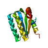
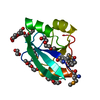
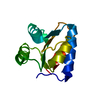
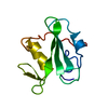
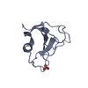
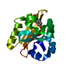
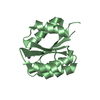
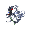
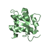
- PDB-1h4x: Structure of the Bacillus Cell Fate Determinant SpoIIAA in the Ph... -
+ Open data

- Basic information
| Entry | Database: PDB / ID: 1h4x | ||||||
|---|---|---|---|---|---|---|---|
| Title | Structure of the Bacillus Cell Fate Determinant SpoIIAA in the Phosphorylated Form | ||||||
 Components Components | ANTI-SIGMA F FACTOR ANTAGONIST | ||||||
 Keywords Keywords | CELL DIFFERENTIATION / PHOSPHORYLATION / SIGMA FACTOR / SPORULATION | ||||||
| Function / homology |  Function and homology information Function and homology informationanti-sigma factor antagonist activity / antisigma factor binding / sporulation resulting in formation of a cellular spore Similarity search - Function | ||||||
| Biological species |  BACILLUS SPHAERICUS (bacteria) BACILLUS SPHAERICUS (bacteria) | ||||||
| Method |  X-RAY DIFFRACTION / SYNCHROTRON / MOLECULAR REPLACEMENT / Resolution: 1.16 Å X-RAY DIFFRACTION / SYNCHROTRON / MOLECULAR REPLACEMENT / Resolution: 1.16 Å | ||||||
 Authors Authors | Seavers, P.R. / Lewis, R.J. / Brannigan, J.A. / Verschueren, K.H.G. / Murshudov, G.N. / Wilkinson, A.J. | ||||||
 Citation Citation | Journal: Structure / Year: 2001 Title: Structure of the Bacillus Cell Fate Determinant Spoiiaa in Phosphorylated and Unphosphorylated Forms Authors: Seavers, P.R. / Lewis, R.J. / Brannigan, J.A. / Verschueren, K.H.G. / Murshudov, G.N. / Wilkinson, A.J. | ||||||
| History |
|
- Structure visualization
| Structure viewer | Molecule: MolmilJmol/JSmol |
|---|
- Downloads & links
-Download
| PDBx/mmCIF format | 1h4x.cif.gz | 68.7 KB | Display | PDBx/mmCIF format |
|---|---|---|---|---|
| PDB format | pdb1h4x.ent.gz | 52 KB | Display | PDB format |
| PDBx/mmJSON format | 1h4x.json.gz | Tree view | PDBx/mmJSON format | |
| Others |  Other downloads Other downloads |
-Validation report
| Arichive directory | https://data.pdbj.org/pub/pdb/validation_reports/h4/1h4xftp://data.pdbj.org/pub/pdb/validation_reports/h4/1h4x | HTTPS FTP |
|---|
-Related structure data


-Links
 PDBj
PDBj
- Assembly
| Deposited unit | 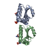
| ||||||||
|---|---|---|---|---|---|---|---|---|---|
| 1 | 
| ||||||||
| 2 | 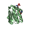
| ||||||||
| Unit cell |
|
-Components
| #1: Protein | Mass: 13205.202 Da / Num. of mol.: 2 Source method: isolated from a genetically manipulated source Source: (gene. exp.) BACILLUS SPHAERICUS (bacteria) / Strain: ATCC14577 / Plasmid: PET28A / Production host: #2: Chemical |   Mass: 122.143 Da / Num. of mol.: 2 / Source method: obtained synthetically / Formula: C4H12NO3 / Comment: pH buffer*YM Mass: 122.143 Da / Num. of mol.: 2 / Source method: obtained synthetically / Formula: C4H12NO3 / Comment: pH buffer*YM#3: Water | ChemComp-HOH / |  Mass: 18.015 Da / Num. of mol.: 426 / Source method: isolated from a natural source / Formula: H2O Mass: 18.015 Da / Num. of mol.: 426 / Source method: isolated from a natural source / Formula: H2OCompound details | IN THE PHOSPHORYL | Has protein modification | Y | Sequence details | THE SEQUENCE SPOIIA FROM BACILLUS SPHAERICUSS STRAIN ATCC14577, IS SIMILAR TO THE SWISSPROT ENTRY ...THE SEQUENCE SPOIIA FROM BACILLUS SPHAERICUS | |
|---|
-Experimental details
-Experiment
| Experiment | Method: X-RAY DIFFRACTION / Number of used crystals: 1 |
|---|
- Sample preparation
| Crystal | Density Matthews: 1.96 Å3/Da / Density % sol: 37.35 % | ||||||||||||||||||||||||||||||||||||||||||||||||
|---|---|---|---|---|---|---|---|---|---|---|---|---|---|---|---|---|---|---|---|---|---|---|---|---|---|---|---|---|---|---|---|---|---|---|---|---|---|---|---|---|---|---|---|---|---|---|---|---|---|
| Crystal grow | pH: 6.5 Details: 30% PEG 5K MME, 200MM (NH4)2SO4, 100MM MES (PH 6.5) | ||||||||||||||||||||||||||||||||||||||||||||||||
| Crystal grow | *PLUS Temperature: 291 K / pH: 8.5 / Method: vapor diffusion, hanging drop / Details: Seavers, P.R., (2001) Acta Crystallogr., D57, 292. | ||||||||||||||||||||||||||||||||||||||||||||||||
| Components of the solutions | *PLUS
|
-Data collection
| Diffraction | Mean temperature: 100 K |
|---|---|
| Diffraction source | Source: SYNCHROTRON / Site: ESRF  / Beamline: ID14-1 / Wavelength: 0.934 / Beamline: ID14-1 / Wavelength: 0.934 |
| Detector | Type: ADSC CCD / Detector: CCD |
| Radiation | Protocol: SINGLE WAVELENGTH / Monochromatic (M) / Laue (L): M / Scattering type: x-ray |
| Radiation wavelength | Wavelength: 0.934 Å / Relative weight: 1 |
| Reflection | Resolution: 1.16→20 Å / Num. obs: 72227 / % possible obs: 97.9 % / Redundancy: 3.8 % / Rmerge(I) obs: 0.068 / Net I/σ(I): 18.9 |
| Reflection shell | Resolution: 1.16→1.2 Å / Redundancy: 2.5 % / Rmerge(I) obs: 0.3 / Mean I/σ(I) obs: 3.72 / % possible all: 85.4 |
| Reflection | *PLUS Num. measured all: 277544 |
| Reflection shell | *PLUS % possible obs: 85.4 % / Rmerge(I) obs: 0.3 |
- Processing
| Software |
| ||||||||||||||||||||||||||||||||||||||||||||||||||||||||||||||||||||||||||||||||||||||||||||||||||||||||||||||||||||||||||||||||||||||||||||||||||||||||||||||||||||||||||||||||||||||
|---|---|---|---|---|---|---|---|---|---|---|---|---|---|---|---|---|---|---|---|---|---|---|---|---|---|---|---|---|---|---|---|---|---|---|---|---|---|---|---|---|---|---|---|---|---|---|---|---|---|---|---|---|---|---|---|---|---|---|---|---|---|---|---|---|---|---|---|---|---|---|---|---|---|---|---|---|---|---|---|---|---|---|---|---|---|---|---|---|---|---|---|---|---|---|---|---|---|---|---|---|---|---|---|---|---|---|---|---|---|---|---|---|---|---|---|---|---|---|---|---|---|---|---|---|---|---|---|---|---|---|---|---|---|---|---|---|---|---|---|---|---|---|---|---|---|---|---|---|---|---|---|---|---|---|---|---|---|---|---|---|---|---|---|---|---|---|---|---|---|---|---|---|---|---|---|---|---|---|---|---|---|---|---|
| Refinement | Method to determine structure: MOLECULAR REPLACEMENT / Resolution: 1.16→19.61 Å / Cor.coef. Fo:Fc: 0.979 / Cor.coef. Fo:Fc free: 0.971 / SU B: 1.218 / SU ML: 0.03 / Cross valid method: THROUGHOUT / ESU R: 0.034 / ESU R Free: 0.035 / Stereochemistry target values: MAXIMUM LIKELIHOOD / Details: HYDROGENS HAVE BEEN ADDED IN THE RIDING POSITIONS
| ||||||||||||||||||||||||||||||||||||||||||||||||||||||||||||||||||||||||||||||||||||||||||||||||||||||||||||||||||||||||||||||||||||||||||||||||||||||||||||||||||||||||||||||||||||||
| Solvent computation | Ion probe radii: 0.8 Å / Shrinkage radii: 0.8 Å / VDW probe radii: 1.4 Å / Solvent model: BABINET MODEL PLUS MASK | ||||||||||||||||||||||||||||||||||||||||||||||||||||||||||||||||||||||||||||||||||||||||||||||||||||||||||||||||||||||||||||||||||||||||||||||||||||||||||||||||||||||||||||||||||||||
| Refinement step | Cycle: LAST / Resolution: 1.16→19.61 Å
| ||||||||||||||||||||||||||||||||||||||||||||||||||||||||||||||||||||||||||||||||||||||||||||||||||||||||||||||||||||||||||||||||||||||||||||||||||||||||||||||||||||||||||||||||||||||
| Refine LS restraints |
|