 Movie
Movie Controller

[English] 日本語
 Yorodumi
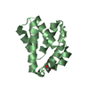
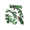
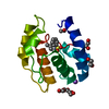
- PDB-2p2e: Crystal structure of a putative Fe-S biosynthesis protein from La... -
+ Open data

- Basic information
| Entry | Database: PDB / ID: 2p2e | ||||||
|---|---|---|---|---|---|---|---|
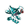
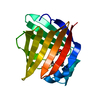
| Title | Crystal structure of a putative Fe-S biosynthesis protein from Lactobacillus salivarius with novel protein fold | ||||||
 Components Components | Putative Fe-S biosynthesis protein | ||||||
 Keywords Keywords | STRUCTURAL GENOMICS / UNKNOWN FUNCTION / hypothetical protein / beta-barrel / Fe-S biosynthesis / 10399l / Protein Structure Initiative / PSI-2 / New York SGX Research Center for Structural Genomics / NYSGXRC | ||||||
| Function / homology | Hypothetical Protein Aq_1857; Chain: A; / HesB-like domain / FeS cluster biogenesis / HesB-like domain superfamily / Iron-sulphur cluster biosynthesis / Sandwich / Mainly Beta / Core domain-containing protein Function and homology information Function and homology information | ||||||
| Biological species |  Lactobacillus salivarius (bacteria) Lactobacillus salivarius (bacteria) | ||||||
| Method |  X-RAY DIFFRACTION / SYNCHROTRON / SAD / Resolution: 2.48 Å X-RAY DIFFRACTION / SYNCHROTRON / SAD / Resolution: 2.48 Å | ||||||
 Authors Authors | Agarwal, R. / Burley, S.K. / Swaminathan, S. / New York SGX Research Center for Structural Genomics (NYSGXRC) | ||||||
 Citation Citation | Journal: To be Published Title: Crystal structure of a putative Fe-S biosynthesis protein from Lactobacillus salivarius with novel protein fold Authors: Agarwal, R. / Burley, S.K. / Swaminathan, S. | ||||||
| History |
|
- Structure visualization
| Structure viewer | Molecule: MolmilJmol/JSmol |
|---|
- Downloads & links
-Download
| PDBx/mmCIF format | 2p2e.cif.gz | 36.6 KB | Display | PDBx/mmCIF format |
|---|---|---|---|---|
| PDB format | pdb2p2e.ent.gz | 25 KB | Display | PDB format |
| PDBx/mmJSON format | 2p2e.json.gz | Tree view | PDBx/mmJSON format | |
| Others |  Other downloads Other downloads |
-Validation report
| Arichive directory | https://data.pdbj.org/pub/pdb/validation_reports/p2/2p2eftp://data.pdbj.org/pub/pdb/validation_reports/p2/2p2e | HTTPS FTP |
|---|
-Related structure data
| Similar structure data | |
|---|---|
| Other databases |


-Links
 PDBj
PDBj- Assembly
| Deposited unit | 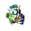
| ||||||||
|---|---|---|---|---|---|---|---|---|---|
| 1 |
| ||||||||
| Unit cell |
|
-Components
| #1: Protein | Mass: 14826.751 Da / Num. of mol.: 1 Source method: isolated from a genetically manipulated source Source: (gene. exp.) Lactobacillus salivarius (bacteria) / Strain: UCC118 / Gene: LSL_1730 / Plasmid: PSGX3(BC) / Production host: |
|---|---|
| #2: Water | ChemComp-HOH /  Mass: 18.015 Da / Num. of mol.: 30 / Source method: isolated from a natural source / Formula: H2O Mass: 18.015 Da / Num. of mol.: 30 / Source method: isolated from a natural source / Formula: H2O |
| Has protein modification | Y |
-Experimental details
-Experiment
| Experiment | Method: X-RAY DIFFRACTION / Number of used crystals: 1 |
|---|
- Sample preparation
| Crystal | Density Matthews: 3.21 Å3/Da / Density % sol: 61.67 % |
|---|---|
| Crystal grow | Temperature: 278 K / Method: vapor diffusion, sitting drop / pH: 4.6 Details: 0.1M Ammonium acetate, 0.1M Sodium acetate, 25% PEG 4000, pH 4.6, VAPOR DIFFUSION, SITTING DROP, temperature 278K |
-Data collection
| Diffraction | Mean temperature: 100 K |
|---|---|
| Diffraction source | Source: SYNCHROTRON / Site: NSLS  / Beamline: X29A / Wavelength: 0.9792 Å / Beamline: X29A / Wavelength: 0.9792 Å |
| Detector | Type: ADSC QUANTUM 315 / Detector: CCD / Date: Mar 2, 2007 / Details: mirrors |
| Radiation | Monochromator: Si-III / Protocol: SINGLE WAVELENGTH / Monochromatic (M) / Laue (L): M / Scattering type: x-ray |
| Radiation wavelength | Wavelength: 0.9792 Å / Relative weight: 1 |
| Reflection | Resolution: 2.48→50 Å / Num. all: 6368 / Num. obs: 6368 / % possible obs: 91.8 % / Observed criterion σ(F): 0 / Observed criterion σ(I): 0 / Redundancy: 16.4 % / Biso Wilson estimate: 30.1 Å2 / Rmerge(I) obs: 0.09 / Net I/σ(I): 7.6 |
| Reflection shell | Resolution: 2.48→2.57 Å / Redundancy: 3 % / Rmerge(I) obs: 0.34 / Mean I/σ(I) obs: 1 / Num. unique all: 352 / % possible all: 52.1 |
- Processing
| Software |
| |||||||||||||||||||||||||
|---|---|---|---|---|---|---|---|---|---|---|---|---|---|---|---|---|---|---|---|---|---|---|---|---|---|---|
| Refinement | Method to determine structure: SAD / Resolution: 2.48→47.29 Å / Rfactor Rfree error: 0.019 / Data cutoff high absF: 45461.85 / Data cutoff low absF: 0 / Isotropic thermal model: RESTRAINED / Cross valid method: THROUGHOUT / σ(F): 0 / Stereochemistry target values: Engh & Huber
| |||||||||||||||||||||||||
| Solvent computation | Solvent model: FLAT MODEL / Bsol: 31.4843 Å2 / ksol: 0.366131 e/Å3 | |||||||||||||||||||||||||
| Displacement parameters | Biso mean: 28.1 Å2
| |||||||||||||||||||||||||
| Refine analyze |
| |||||||||||||||||||||||||
| Refinement step | Cycle: LAST / Resolution: 2.48→47.29 Å
| |||||||||||||||||||||||||
| Refine LS restraints |
| |||||||||||||||||||||||||
| LS refinement shell | Resolution: 2.48→2.64 Å / Rfactor Rfree error: 0.068 / Total num. of bins used: 6
| |||||||||||||||||||||||||
| Xplor file |
|
