 Movie
Movie Controller


+ Open data

- Basic information
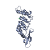
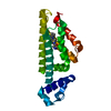
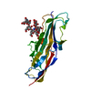
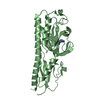
| Entry | Database: PDB / ID: 3hlr | ||||||
|---|---|---|---|---|---|---|---|
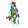
| Title | Donor strand complemented FaeG of F4ad fimbriae | ||||||
 Components Components | K88 fimbrial protein AD | ||||||
 Keywords Keywords | CELL ADHESION / immunoglobuline like fold / Fimbrium | ||||||
| Function / homology | Fimbrial, major/minor subunit / Fimbrial, major and minor subunit / pilus / cell adhesion / K88 fimbrial protein AD Function and homology information Function and homology information | ||||||
| Biological species |  | ||||||
| Method |  X-RAY DIFFRACTION / SYNCHROTRON / MOLECULAR REPLACEMENT / Resolution: 2.3 Å X-RAY DIFFRACTION / SYNCHROTRON / MOLECULAR REPLACEMENT / Resolution: 2.3 Å | ||||||
 Authors Authors | Van Molle, I. / Moonens, K. / Garcia-Pino, A. / Buts, L. / Bouckaert, J. / De Greve, H. | ||||||
 Citation Citation | Journal: J.Mol.Biol. / Year: 2009 Title: Structural and thermodynamic characterization of pre- and postpolymerization states in the F4 fimbrial subunit FaeG Authors: Van Molle, I. / Moonens, K. / Garcia-Pino, A. / Buts, L. / De Kerpel, M. / Wyns, L. / Bouckaert, J. / De Greve, H. | ||||||
| History |
|
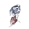
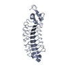
- Structure visualization
| Structure viewer | Molecule: MolmilJmol/JSmol |
|---|
- Downloads & links
-Download
| PDBx/mmCIF format | 3hlr.cif.gz | 65.2 KB | Display | PDBx/mmCIF format |
|---|---|---|---|---|
| PDB format | pdb3hlr.ent.gz | 46.8 KB | Display | PDB format |
| PDBx/mmJSON format | 3hlr.json.gz | Tree view | PDBx/mmJSON format | |
| Others |  Other downloads Other downloads |
-Validation report
| Summary document | 3hlr_validation.pdf.gz | 422.6 KB | Display | wwPDB validaton report |
|---|---|---|---|---|
| Full document | 3hlr_full_validation.pdf.gz | 430.5 KB | Display | |
| Data in XML | 3hlr_validation.xml.gz | 14.4 KB | Display | |
| Data in CIF | 3hlr_validation.cif.gz | 20.1 KB | Display | |
| Arichive directory | https://data.pdbj.org/pub/pdb/validation_reports/hl/3hlrftp://data.pdbj.org/pub/pdb/validation_reports/hl/3hlr | HTTPS FTP |
-Related structure data
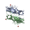
| Related structure data | 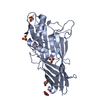 3geaSC  3gewC 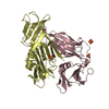 3gfuC  3gghC C: citing same article ( S: Starting model for refinement |
|---|---|
| Similar structure data |


-Links
 PDBj
PDBj- Assembly
| Deposited unit | 
| ||||||||
|---|---|---|---|---|---|---|---|---|---|
| 1 |
| ||||||||
| Unit cell |
|
-Components
| #1: Protein | Mass: 28344.375 Da / Num. of mol.: 1 / Fragment: FaeGntd/dsc2 Source method: isolated from a genetically manipulated source Details: The sequence consists of MG, UNP residues 43-285 (Uniprot 14191), a linker of 10 GLYs, and UNP residues 22-42 (Uniprot P14191). Source: (gene. exp.) |
|---|---|
| #2: Water | ChemComp-HOH /  Mass: 18.015 Da / Num. of mol.: 194 / Source method: isolated from a natural source / Formula: H2O Mass: 18.015 Da / Num. of mol.: 194 / Source method: isolated from a natural source / Formula: H2O |
| Sequence details | THE CONFLICT BASED ON REFERENCE 2 OF DATABASE FAEG3_ECOLX (UNIPROTKB/SWISS-PROT P14191). N19S (UNP ...THE CONFLICT BASED ON REFERENCE 2 OF DATABASE FAEG3_ECOLX (UNIPROTKB/SWISS-PROT P14191). N19S (UNP RESIDUE 59) IS CONFLICT OF FAEG3_ECOLX. |
-Experimental details
-Experiment
| Experiment | Method: X-RAY DIFFRACTION / Number of used crystals: 1 |
|---|
- Sample preparation
| Crystal | Density Matthews: 1.96 Å3/Da / Density % sol: 37.17 % |
|---|---|
| Crystal grow | Temperature: 298 K / Method: vapor diffusion, hanging drop / pH: 6.5 Details: 0.1M Na cacodylate pH 6.5, 0.2M ammonium acetate, 20% PEG, VAPOR DIFFUSION, HANGING DROP, temperature 298K |
-Data collection
| Diffraction | Mean temperature: 100 K |
|---|---|
| Diffraction source | Source: SYNCHROTRON / Site: SOLEIL  / Beamline: PROXIMA 1 / Wavelength: 0.9801 Å / Beamline: PROXIMA 1 / Wavelength: 0.9801 Å |
| Detector | Type: MAR555 FLAT PANEL / Detector: IMAGE PLATE / Date: Jan 31, 2009 |
| Radiation | Monochromator: Graphite / Protocol: SINGLE WAVELENGTH / Monochromatic (M) / Laue (L): M / Scattering type: x-ray |
| Radiation wavelength | Wavelength: 0.9801 Å / Relative weight: 1 |
| Reflection | Resolution: 2.3→35 Å / Num. all: 64572 / Num. obs: 64572 / % possible obs: 100 % / Observed criterion σ(F): 0 / Observed criterion σ(I): 0 / Biso Wilson estimate: 22.28 Å2 / Rmerge(I) obs: 0.1572 / Net I/σ(I): 2.97 |
| Reflection shell | Resolution: 2.3→2.32 Å / Rmerge(I) obs: 0.32 / Mean I/σ(I) obs: 1.97 / % possible all: 97 |
- Processing
| Software |
| ||||||||||||||||||||||||||||
|---|---|---|---|---|---|---|---|---|---|---|---|---|---|---|---|---|---|---|---|---|---|---|---|---|---|---|---|---|---|
| Refinement | Method to determine structure: MOLECULAR REPLACEMENT Starting model: PDB ENTRY 3GEA Resolution: 2.3→29.162 Å / SU ML: 0.4 / Cross valid method: THROUGHOUT / σ(F): 2 / σ(I): 0 / Stereochemistry target values: ML
| ||||||||||||||||||||||||||||
| Solvent computation | Shrinkage radii: 0.9 Å / VDW probe radii: 1.11 Å / Solvent model: FLAT BULK SOLVENT MODEL / Bsol: 35.961 Å2 / ksol: 0.33 e/Å3 | ||||||||||||||||||||||||||||
| Displacement parameters | Biso mean: 24.204 Å2
| ||||||||||||||||||||||||||||
| Refinement step | Cycle: LAST / Resolution: 2.3→29.162 Å
| ||||||||||||||||||||||||||||
| Refine LS restraints |
| ||||||||||||||||||||||||||||
| LS refinement shell |
|
