 Movie
Movie Controller

[English] 日本語
 Yorodumi
- PDB-3hjm: Crystal structure of human Glutathione Transferase Pi Y108V mutant -
+ Open data

- Basic information
| Entry | Database: PDB / ID: 3hjm | ||||||
|---|---|---|---|---|---|---|---|
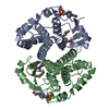
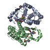
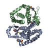
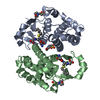
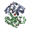
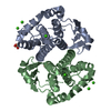
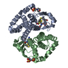
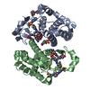
| Title | Crystal structure of human Glutathione Transferase Pi Y108V mutant | ||||||
 Components Components | Glutathione S-transferase P | ||||||
 Keywords Keywords | TRANSFERASE / GLUTATHIONE / DETOXIFICATION / MUTANT / Polymorphism | ||||||
| Function / homology |  Function and homology information Function and homology informationnitric oxide storage / S-nitrosoglutathione binding / TRAF2-GSTP1 complex / negative regulation of smooth muscle cell chemotaxis / negative regulation of leukocyte proliferation / dinitrosyl-iron complex binding / common myeloid progenitor cell proliferation / hepoxilin biosynthetic process / cellular response to cell-matrix adhesion / glutathione derivative biosynthetic process ...nitric oxide storage / S-nitrosoglutathione binding / TRAF2-GSTP1 complex / negative regulation of smooth muscle cell chemotaxis / negative regulation of leukocyte proliferation / dinitrosyl-iron complex binding / common myeloid progenitor cell proliferation / hepoxilin biosynthetic process / cellular response to cell-matrix adhesion / glutathione derivative biosynthetic process / response to L-ascorbic acid / linoleic acid metabolic process / Glutathione conjugation / nitric oxide binding / negative regulation of monocyte chemotactic protein-1 production / JUN kinase binding / glutathione peroxidase activity / Paracetamol ADME / oligodendrocyte development / negative regulation of stress-activated MAPK cascade / negative regulation of JNK cascade / cellular response to glucocorticoid stimulus / prostaglandin metabolic process / negative regulation of interleukin-1 beta production / regulation of stress-activated MAPK cascade / Detoxification of Reactive Oxygen Species / negative regulation of acute inflammatory response / glutathione transferase / negative regulation of ferroptosis / negative regulation of vascular associated smooth muscle cell proliferation / response to amino acid / glutathione transferase activity / animal organ regeneration / negative regulation of tumor necrosis factor production / protein serine/threonine kinase inhibitor activity / negative regulation of tumor necrosis factor-mediated signaling pathway / regulation of ERK1 and ERK2 cascade / toxic substance binding / negative regulation of fibroblast proliferation / positive regulation of superoxide anion generation / negative regulation of MAPK cascade / xenobiotic metabolic process / cellular response to epidermal growth factor stimulus / fatty acid binding / negative regulation of extrinsic apoptotic signaling pathway / response to reactive oxygen species / central nervous system development / negative regulation of canonical NF-kappaB signal transduction / glutathione metabolic process / negative regulation of ERK1 and ERK2 cascade / cellular response to insulin stimulus / response to estradiol / cellular response to lipopolysaccharide / secretory granule lumen / vesicle / ficolin-1-rich granule lumen / response to ethanol / Neutrophil degranulation / negative regulation of apoptotic process / negative regulation of transcription by RNA polymerase II / mitochondrion / : / extracellular exosome / extracellular region / nucleus / cytoplasm / cytosol Similarity search - Function | ||||||
| Biological species |  Homo sapiens (human) Homo sapiens (human) | ||||||
| Method |  X-RAY DIFFRACTION / MOLECULAR REPLACEMENT / Resolution: 2.1 Å X-RAY DIFFRACTION / MOLECULAR REPLACEMENT / Resolution: 2.1 Å | ||||||
 Authors Authors | Parker, L.J. | ||||||
 Citation Citation | Journal: Protein Sci. / Year: 2009 Title: Influence of the H-site residue 108 on human glutathione transferase P1-1 ligand binding: structure-thermodynamic relationships and thermal stability. Authors: Quesada-Soriano, I. / Parker, L.J. / Primavera, A. / Casas-Solvas, J.M. / Vargas-Berenguel, A. / Baron, C. / Morton, C.J. / Mazzetti, A.P. / Lo Bello, M. / Parker, M.W. / Garcia-Fuentes, L. | ||||||
| History |
|
- Structure visualization
| Structure viewer | Molecule: MolmilJmol/JSmol |
|---|
- Downloads & links
-Download
| PDBx/mmCIF format | 3hjm.cif.gz | 186.3 KB | Display | PDBx/mmCIF format |
|---|---|---|---|---|
| PDB format | pdb3hjm.ent.gz | 147.7 KB | Display | PDB format |
| PDBx/mmJSON format | 3hjm.json.gz | Tree view | PDBx/mmJSON format | |
| Others |  Other downloads Other downloads |
-Validation report
| Arichive directory | https://data.pdbj.org/pub/pdb/validation_reports/hj/3hjmftp://data.pdbj.org/pub/pdb/validation_reports/hj/3hjm | HTTPS FTP |
|---|
-Related structure data
| Related structure data |  3hjoC  3hkrC  3csiS C: citing same article ( S: Starting model for refinement |
|---|---|
| Similar structure data |

-Links
 PDBj
PDBj


- Assembly
| Deposited unit | 
| ||||||||
|---|---|---|---|---|---|---|---|---|---|
| 1 | 
| ||||||||
| 2 | 
| ||||||||
| Unit cell |
|
-Components
-Protein , 1 types, 4 molecules ABCD
| #1: Protein | Mass: 23182.527 Da / Num. of mol.: 4 / Mutation: Y108V Source method: isolated from a genetically manipulated source Source: (gene. exp.) Homo sapiens (human) / Gene: GSTP1 / Plasmid: pSE420 / Production host:  |
|---|
-Non-polymers , 6 types, 638 molecules 


| #2: Chemical |  Mass: 195.237 Da / Num. of mol.: 3 / Source method: obtained synthetically / Formula: C6H13NO4S / Comment: pH buffer*YM Mass: 195.237 Da / Num. of mol.: 3 / Source method: obtained synthetically / Formula: C6H13NO4S / Comment: pH buffer*YM#3: Chemical | ChemComp-CA /  Mass: 40.078 Da / Num. of mol.: 5 / Source method: obtained synthetically / Formula: Ca Mass: 40.078 Da / Num. of mol.: 5 / Source method: obtained synthetically / Formula: Ca#4: Chemical | ChemComp-CO3 /  Mass: 60.009 Da / Num. of mol.: 5 / Source method: obtained synthetically / Formula: CO3 Mass: 60.009 Da / Num. of mol.: 5 / Source method: obtained synthetically / Formula: CO3#5: Chemical |  Mass: 35.453 Da / Num. of mol.: 3 / Source method: obtained synthetically / Formula: Cl Mass: 35.453 Da / Num. of mol.: 3 / Source method: obtained synthetically / Formula: Cl#6: Chemical | ChemComp-PO4 / |  Mass: 94.971 Da / Num. of mol.: 1 / Source method: obtained synthetically / Formula: PO4 Mass: 94.971 Da / Num. of mol.: 1 / Source method: obtained synthetically / Formula: PO4#7: Water | ChemComp-HOH / | Mass: 18.015 Da / Num. of mol.: 621 / Source method: isolated from a natural source / Formula: H2O |
|---|
-Experimental details
-Experiment
| Experiment | Method: X-RAY DIFFRACTION / Number of used crystals: 1 |
|---|
- Sample preparation
| Crystal | Density Matthews: 2.53 Å3/Da / Density % sol: 51.45 % / Mosaicity: 0.635 ° |
|---|---|
| Crystal grow | Temperature: 298 K / Method: vapor diffusion, hanging drop / pH: 5.4 Details: 270mM Calcium Acetate, 20% PEG8000, 100mM MES, pH5.4, VAPOR DIFFUSION, HANGING DROP, temperature 298K |
-Data collection
| Diffraction | Mean temperature: 100 K |
|---|---|
| Diffraction source | Source: ROTATING ANODE / Type: RIGAKU MICROMAX-007 HF / Wavelength: 1.54 Å |
| Detector | Type: RIGAKU RAXIS IV++ / Detector: IMAGE PLATE / Date: Aug 15, 2008 / Details: AXCO |
| Radiation | Monochromator: AXCO Mirrors / Protocol: SINGLE WAVELENGTH / Monochromatic (M) / Laue (L): M / Scattering type: x-ray |
| Radiation wavelength | Wavelength: 1.54 Å / Relative weight: 1 |
| Reflection | Resolution: 2.1→54.55 Å / Num. obs: 53439 / % possible obs: 98.8 % / Redundancy: 5.7 % / Biso Wilson estimate: 21.99 Å2 / Rmerge(I) obs: 0.11 / Rsym value: 0.11 / Net I/σ(I): 6.26 |
| Reflection shell | Resolution: 2.1→2.21 Å / Redundancy: 4.8 % / Rmerge(I) obs: 0.41 / Mean I/σ(I) obs: 1.8 / Num. measured all: 34588 / Num. unique all: 7215 / Rsym value: 0.41 / % possible all: 92.1 |
- Processing
| Software |
| |||||||||||||||||||||||||||||||||||||||||||||||||||||||||||||||||||||||||||||||||||||||||||||||
|---|---|---|---|---|---|---|---|---|---|---|---|---|---|---|---|---|---|---|---|---|---|---|---|---|---|---|---|---|---|---|---|---|---|---|---|---|---|---|---|---|---|---|---|---|---|---|---|---|---|---|---|---|---|---|---|---|---|---|---|---|---|---|---|---|---|---|---|---|---|---|---|---|---|---|---|---|---|---|---|---|---|---|---|---|---|---|---|---|---|---|---|---|---|---|---|---|
| Refinement | Method to determine structure: MOLECULAR REPLACEMENT Starting model: PDB ENTRY 3CSI Resolution: 2.1→54.55 Å / Cor.coef. Fo:Fc: 0.957 / Cor.coef. Fo:Fc free: 0.933 / WRfactor Rfree: 0.2 / WRfactor Rwork: 0.16 / Occupancy max: 1 / Occupancy min: 0.5 / FOM work R set: 0.834 / SU B: 5.108 / SU ML: 0.135 / SU R Cruickshank DPI: 0.218 / SU Rfree: 0.185 / Cross valid method: THROUGHOUT / σ(F): 0 / ESU R: 0.218 / ESU R Free: 0.185 / Stereochemistry target values: MAXIMUM LIKELIHOOD / Details: HYDROGENS HAVE BEEN ADDED IN THE RIDING POSITIONS
| |||||||||||||||||||||||||||||||||||||||||||||||||||||||||||||||||||||||||||||||||||||||||||||||
| Solvent computation | Ion probe radii: 0.8 Å / Shrinkage radii: 0.8 Å / VDW probe radii: 1.2 Å / Solvent model: MASK | |||||||||||||||||||||||||||||||||||||||||||||||||||||||||||||||||||||||||||||||||||||||||||||||
| Displacement parameters | Biso max: 53.88 Å2 / Biso mean: 21.361 Å2 / Biso min: 2.75 Å2
| |||||||||||||||||||||||||||||||||||||||||||||||||||||||||||||||||||||||||||||||||||||||||||||||
| Refinement step | Cycle: LAST / Resolution: 2.1→54.55 Å
| |||||||||||||||||||||||||||||||||||||||||||||||||||||||||||||||||||||||||||||||||||||||||||||||
| Refine LS restraints |
| |||||||||||||||||||||||||||||||||||||||||||||||||||||||||||||||||||||||||||||||||||||||||||||||
| LS refinement shell | Resolution: 2.1→2.16 Å / Total num. of bins used: 20
|
