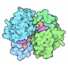
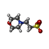
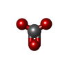
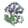
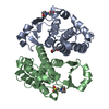
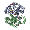
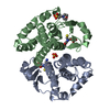
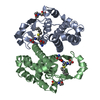
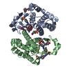
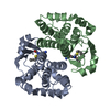
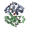
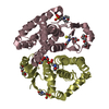
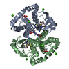
Entry Database : PDB / ID : 3hkrTitle Crystal Structure of Glutathione Transferase Pi Y108V Mutant Glutathione S-transferase P Keywords / / Function / homology Function Domain/homology Component
/ / / / / / / / / / / / / / / / / / / / / / / / / / / / / / / / / / / / / / / / / / / / / / / / / / / / / / / / / / / / / / / / / / / / / / / / / / / / / / / / / / / / / / / / / / / / / / / / / / / / / / Biological species Homo sapiens (human)Method / / / Resolution : 1.8 Å Authors Parker, L.J. Journal : Protein Sci. / Year : 2009Title : Influence of the H-site residue 108 on human glutathione transferase P1-1 ligand binding: structure-thermodynamic relationships and thermal stability.Authors : Quesada-Soriano, I. / Parker, L.J. / Primavera, A. / Casas-Solvas, J.M. / Vargas-Berenguel, A. / Baron, C. / Morton, C.J. / Mazzetti, A.P. / Lo Bello, M. / Parker, M.W. / Garcia-Fuentes, L. History Deposition May 25, 2009 Deposition site / Processing site Revision 1.0 Sep 22, 2009 Provider / Type Revision 1.1 Jul 13, 2011 Group Revision 1.2 Nov 10, 2021 Group / Derived calculationsCategory database_2 / pdbx_struct_conn_angle ... database_2 / pdbx_struct_conn_angle / struct_conn / struct_ref_seq_dif / struct_site Item _database_2.pdbx_DOI / _database_2.pdbx_database_accession ... _database_2.pdbx_DOI / _database_2.pdbx_database_accession / _pdbx_struct_conn_angle.ptnr1_auth_asym_id / _pdbx_struct_conn_angle.ptnr1_auth_comp_id / _pdbx_struct_conn_angle.ptnr1_auth_seq_id / _pdbx_struct_conn_angle.ptnr1_label_asym_id / _pdbx_struct_conn_angle.ptnr1_label_atom_id / _pdbx_struct_conn_angle.ptnr1_label_comp_id / _pdbx_struct_conn_angle.ptnr1_label_seq_id / _pdbx_struct_conn_angle.ptnr3_auth_asym_id / _pdbx_struct_conn_angle.ptnr3_auth_comp_id / _pdbx_struct_conn_angle.ptnr3_auth_seq_id / _pdbx_struct_conn_angle.ptnr3_label_asym_id / _pdbx_struct_conn_angle.ptnr3_label_atom_id / _pdbx_struct_conn_angle.ptnr3_label_comp_id / _pdbx_struct_conn_angle.ptnr3_label_seq_id / _pdbx_struct_conn_angle.value / _struct_conn.pdbx_dist_value / _struct_conn.ptnr1_auth_asym_id / _struct_conn.ptnr1_auth_comp_id / _struct_conn.ptnr1_auth_seq_id / _struct_conn.ptnr1_label_asym_id / _struct_conn.ptnr1_label_atom_id / _struct_conn.ptnr1_label_comp_id / _struct_conn.ptnr1_label_seq_id / _struct_conn.ptnr2_auth_asym_id / _struct_conn.ptnr2_auth_comp_id / _struct_conn.ptnr2_auth_seq_id / _struct_conn.ptnr2_label_asym_id / _struct_conn.ptnr2_label_atom_id / _struct_conn.ptnr2_label_comp_id / _struct_conn.ptnr2_label_seq_id / _struct_ref_seq_dif.details / _struct_site.pdbx_auth_asym_id / _struct_site.pdbx_auth_comp_id / _struct_site.pdbx_auth_seq_id Revision 1.3 Nov 1, 2023 Group / Refinement descriptionCategory / chem_comp_bond / pdbx_initial_refinement_model
Show all Show less
 Open data
 Basic information
 Components
 Keywords
 Function and homology information
 Homo sapiens (human)
 X-RAY DIFFRACTION /
  SYNCHROTRON /
  MOLECULAR REPLACEMENT / Resolution: 1.8 Å
  Authors
 Citation
 Journal: Protein Sci. / Year: 2009
 Structure visualization
 Molmil
 Jmol/JSmol
 Downloads & links
 Download
 3hkr.cif.gz
 PDBx/mmCIF format
 pdb3hkr.ent.gz
 PDB format
 3hkr.json.gz
 PDBx/mmJSON format
 Other downloads
 https://data.pdbj.org/pub/pdb/validation_reports/hk/3hkr
 ftp://data.pdbj.org/pub/pdb/validation_reports/hk/3hkr
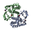
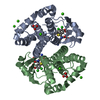
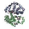
 Links
Links Assembly
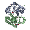
 Components
 Homo sapiens (human) / Gene: GSTP1 / Plasmid: PSE420 / Production host:
 X-RAY DIFFRACTION / Number of used crystals: 1
  Sample preparation
 SYNCHROTRON / Site:
  Australian Synchrotron
  / Beamline: MX1 / Wavelength: 0.96 Å
 Processing
 MOLECULAR REPLACEMENT
 Movie
Movie Controller





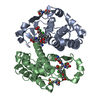
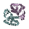
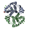
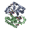
 PDBj
PDBj