 Movie
Movie Controller

[English] 日本語
 Yorodumi
- PDB-3h6j: Crystal structure of a putative neuraminidase from Pseudomonas ae... -
+ Open data

- Basic information
| Entry | Database: PDB / ID: 3h6j | ||||||
|---|---|---|---|---|---|---|---|
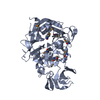
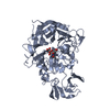
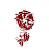
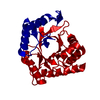
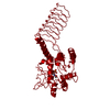
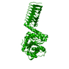
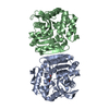
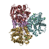
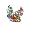
| Title | Crystal structure of a putative neuraminidase from Pseudomonas aeruginosa | ||||||
 Components Components | Neuraminidase | ||||||
 Keywords Keywords | HYDROLASE / SIX-BLADED BETA-PROPELLER / CELL WALL / GLYCOSIDASE / PEPTIDOGLYCAN-ANCHOR / SECRETED | ||||||
| Function / homology |  Function and homology information Function and homology informationganglioside catabolic process / oligosaccharide catabolic process / exo-alpha-sialidase / exo-alpha-sialidase activity / membrane / cytoplasm Similarity search - Function | ||||||
| Biological species |   Pseudomonas aeruginosa (bacteria) Pseudomonas aeruginosa (bacteria) | ||||||
| Method |  X-RAY DIFFRACTION / SYNCHROTRON / SAD / Resolution: 1.6 Å X-RAY DIFFRACTION / SYNCHROTRON / SAD / Resolution: 1.6 Å | ||||||
 Authors Authors | Hsiao, Y.-S. / Tong, L. | ||||||
 Citation Citation | Journal: Biochem.Biophys.Res.Commun. / Year: 2009 Title: Crystal structures of respiratory pathogen neuraminidases Authors: Hsiao, Y.-S. / Parker, D. / Ratner, A.J. / Prince, A. / Tong, L. | ||||||
| History |
|
- Structure visualization
| Structure viewer | Molecule: MolmilJmol/JSmol |
|---|
- Downloads & links
-Download
| PDBx/mmCIF format | 3h6j.cif.gz | 117.2 KB | Display | PDBx/mmCIF format |
|---|---|---|---|---|
| PDB format | pdb3h6j.ent.gz | 88.4 KB | Display | PDB format |
| PDBx/mmJSON format | 3h6j.json.gz | Tree view | PDBx/mmJSON format | |
| Others |  Other downloads Other downloads |
-Validation report
| Arichive directory | https://data.pdbj.org/pub/pdb/validation_reports/h6/3h6jftp://data.pdbj.org/pub/pdb/validation_reports/h6/3h6j | HTTPS FTP |
|---|
-Related structure data


-Links
 PDBj
PDBj

- Assembly
| Deposited unit | 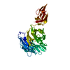
| ||||||||
|---|---|---|---|---|---|---|---|---|---|
| 1 | 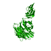
| ||||||||
| Unit cell |
|
-Components
| #1: Protein | Mass: 47157.980 Da / Num. of mol.: 1 Source method: isolated from a genetically manipulated source Source: (gene. exp.) Pseudomonas aeruginosa (bacteria) / Gene: PA2794 / Production host: |
|---|---|
| #2: Water | ChemComp-HOH /  Mass: 18.015 Da / Num. of mol.: 908 / Source method: isolated from a natural source / Formula: H2O Mass: 18.015 Da / Num. of mol.: 908 / Source method: isolated from a natural source / Formula: H2O |
-Experimental details
-Experiment
| Experiment | Method: X-RAY DIFFRACTION / Number of used crystals: 1 |
|---|
- Sample preparation
| Crystal | Density Matthews: 3.56 Å3/Da / Density % sol: 65.48 % |
|---|---|
| Crystal grow | Temperature: 294 K / Method: vapor diffusion, sitting drop / pH: 7 Details: 100 mM Hepes, 5% Tacsimate, 7% (w/v) PEGMME 5000, pH 7.0, VAPOR DIFFUSION, SITTING DROP, temperature 294K |
-Data collection
| Diffraction | Mean temperature: 100 K |
|---|---|
| Diffraction source | Source: SYNCHROTRON / Site: NSLS  / Beamline: X4A / Wavelength: 0.9798 Å / Beamline: X4A / Wavelength: 0.9798 Å |
| Detector | Type: ADSC QUANTUM 4 / Detector: CCD / Date: Apr 23, 2006 / Details: Mirrors |
| Radiation | Monochromator: SI(111) / Protocol: SINGLE WAVELENGTH / Monochromatic (M) / Laue (L): M / Scattering type: x-ray |
| Radiation wavelength | Wavelength: 0.9798 Å / Relative weight: 1 |
| Reflection | Resolution: 1.6→30 Å / Num. obs: 88080 / % possible obs: 99.4 % / Observed criterion σ(F): 0 / Observed criterion σ(I): 0 / Redundancy: 4.3 % / Biso Wilson estimate: 12.9 Å2 / Rmerge(I) obs: 0.088 / Net I/σ(I): 11.8493 |
| Reflection shell | Resolution: 1.6→1.66 Å / Redundancy: 3.9 % / Rmerge(I) obs: 0.365 / Mean I/σ(I) obs: 3.299 / Num. unique all: 8684 / % possible all: 98.9 |
- Processing
| Software |
| ||||||||||||||||||||||||||||||||||||
|---|---|---|---|---|---|---|---|---|---|---|---|---|---|---|---|---|---|---|---|---|---|---|---|---|---|---|---|---|---|---|---|---|---|---|---|---|---|
| Refinement | Method to determine structure: SAD / Resolution: 1.6→26.94 Å / Rfactor Rfree error: 0.002 / Data cutoff high absF: 439616.95 / Data cutoff low absF: 0 / Isotropic thermal model: RESTRAINED / Cross valid method: THROUGHOUT / σ(F): 0 / σ(I): 0 / Stereochemistry target values: Engh & Huber
| ||||||||||||||||||||||||||||||||||||
| Solvent computation | Solvent model: FLAT MODEL / Bsol: 61.7778 Å2 / ksol: 0.348456 e/Å3 | ||||||||||||||||||||||||||||||||||||
| Displacement parameters | Biso mean: 17.9 Å2
| ||||||||||||||||||||||||||||||||||||
| Refine analyze |
| ||||||||||||||||||||||||||||||||||||
| Refinement step | Cycle: LAST / Resolution: 1.6→26.94 Å
| ||||||||||||||||||||||||||||||||||||
| Refine LS restraints |
| ||||||||||||||||||||||||||||||||||||
| LS refinement shell | Resolution: 1.6→1.66 Å / Rfactor Rfree error: 0.01 / Total num. of bins used: 10
| ||||||||||||||||||||||||||||||||||||
| Xplor file |
|
