 Movie
Movie Controller

[English] 日本語
 Yorodumi
- PDB-2qkx: N-acetyl glucosamine 1-phosphate uridyltransferase from Mycobacte... -
+ Open data

- Basic information
| Entry | Database: PDB / ID: 2qkx | |||||||||
|---|---|---|---|---|---|---|---|---|---|---|
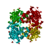
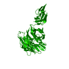
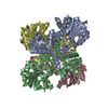
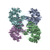
| Title | N-acetyl glucosamine 1-phosphate uridyltransferase from Mycobacterium tuberculosis complex with N-acetyl glucosamine 1-phosphate | |||||||||
 Components Components | Bifunctional protein glmU | |||||||||
 Keywords Keywords | TRANSFERASE / rossmann / beta-helix / substrate complex / Structural Genomics / TB Structural Genomics Consortium / TBSGC | |||||||||
| Function / homology |  Function and homology information Function and homology informationentry of bacterium into host cell / uridylyltransferase activity / adhesion of symbiont to host cell / glucosamine-1-phosphate N-acetyltransferase / glucosamine-1-phosphate N-acetyltransferase activity / UDP-N-acetylglucosamine diphosphorylase / UDP-N-acetylglucosamine diphosphorylase activity / UDP-N-acetylglucosamine biosynthetic process / lipid A biosynthetic process / peptidoglycan biosynthetic process ...entry of bacterium into host cell / uridylyltransferase activity / adhesion of symbiont to host cell / glucosamine-1-phosphate N-acetyltransferase / glucosamine-1-phosphate N-acetyltransferase activity / UDP-N-acetylglucosamine diphosphorylase / UDP-N-acetylglucosamine diphosphorylase activity / UDP-N-acetylglucosamine biosynthetic process / lipid A biosynthetic process / peptidoglycan biosynthetic process / cell wall organization / cell morphogenesis / regulation of cell shape / magnesium ion binding / membrane / cytoplasm / cytosol Similarity search - Function | |||||||||
| Biological species |   Mycobacterium tuberculosis (bacteria) Mycobacterium tuberculosis (bacteria) | |||||||||
| Method |  X-RAY DIFFRACTION / MOLECULAR REPLACEMENT / Resolution: 2.75 Å X-RAY DIFFRACTION / MOLECULAR REPLACEMENT / Resolution: 2.75 Å | |||||||||
 Authors Authors | Zhang, Z. / Squire, C.J. / Baker, E.N. / TB Structural Genomics Consortium (TBSGC) | |||||||||
 Citation Citation | Journal: Acta Crystallogr.,Sect.D / Year: 2009 Title: Structure and function of GlmU from Mycobacterium tuberculosis. Authors: Zhang, Z. / Bulloch, E.M. / Bunker, R.D. / Baker, E.N. / Squire, C.J. | |||||||||
| History |
|
- Structure visualization
| Structure viewer | Molecule: MolmilJmol/JSmol |
|---|
- Downloads & links
-Download
| PDBx/mmCIF format | 2qkx.cif.gz | 83.7 KB | Display | PDBx/mmCIF format |
|---|---|---|---|---|
| PDB format | pdb2qkx.ent.gz | 62.5 KB | Display | PDB format |
| PDBx/mmJSON format | 2qkx.json.gz | Tree view | PDBx/mmJSON format | |
| Others |  Other downloads Other downloads |
-Validation report
| Arichive directory | https://data.pdbj.org/pub/pdb/validation_reports/qk/2qkxftp://data.pdbj.org/pub/pdb/validation_reports/qk/2qkx | HTTPS FTP |
|---|
-Related structure data
| Related structure data |  3d8vC  3d98C  1hm9S S: Starting model for refinement C: citing same article ( |
|---|---|
| Similar structure data |


-Links
 PDBj
PDBj


- Assembly
| Deposited unit | 
| ||||||||
|---|---|---|---|---|---|---|---|---|---|
| 1 | 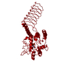
| ||||||||
| Unit cell |
| ||||||||
| Details | The biological assembly is a trimer generated by the 3-fold axis: -x+y,-x,z and -y,x-y,z. |
-Components
| #1: Protein | Mass: 40761.758 Da / Num. of mol.: 1 Source method: isolated from a genetically manipulated source Source: (gene. exp.) Mycobacterium tuberculosis (bacteria) / Strain: H37Rv / Gene: glmU, Rv1018c, MT1046 / Plasmid: pDEST17 / Production host: References: UniProt: P96382, UniProt: P9WMN3*PLUS, glucosamine-1-phosphate N-acetyltransferase |
|---|---|
| #2: Sugar | ChemComp-GN1 /   Type: D-saccharide / Mass: 301.188 Da / Num. of mol.: 1 Type: D-saccharide / Mass: 301.188 Da / Num. of mol.: 1Source method: isolated from a genetically manipulated source Formula: C8H16NO9P |
| #3: Water | ChemComp-HOH /  Mass: 18.015 Da / Num. of mol.: 39 / Source method: isolated from a natural source / Formula: H2O Mass: 18.015 Da / Num. of mol.: 39 / Source method: isolated from a natural source / Formula: H2O |
-Experimental details
-Experiment
| Experiment | Method: X-RAY DIFFRACTION / Number of used crystals: 1 |
|---|
- Sample preparation
| Crystal | Density Matthews: 3.02 Å3/Da / Density % sol: 59.3 % |
|---|---|
| Crystal grow | Temperature: 291 K / Method: vapor diffusion, sitting drop / pH: 7.5 Details: 0.2M Lithium nitrate, 20% PEG 3350, pH 7.5, VAPOR DIFFUSION, SITTING DROP, temperature 291K |
-Data collection
| Diffraction | Mean temperature: 110 K |
|---|---|
| Diffraction source | Source: ROTATING ANODE / Type: RIGAKU MICROMAX-007 / Wavelength: 1.5418 Å |
| Detector | Type: MAR scanner 345 mm plate / Detector: IMAGE PLATE / Date: Jun 30, 2007 / Details: osmic |
| Radiation | Monochromator: osmic / Protocol: SINGLE WAVELENGTH / Monochromatic (M) / Laue (L): M / Scattering type: x-ray |
| Radiation wavelength | Wavelength: 1.5418 Å / Relative weight: 1 |
| Reflection | Resolution: 2.75→42.33 Å / Num. all: 13239 / Num. obs: 13239 / % possible obs: 100 % / Observed criterion σ(F): 0 / Observed criterion σ(I): 0 / Redundancy: 6.3 % / Rmerge(I) obs: 0.151 / Net I/σ(I): 13.2 |
| Reflection shell | Resolution: 2.75→2.9 Å / Redundancy: 6.3 % / Rmerge(I) obs: 0.623 / Mean I/σ(I) obs: 3 / Num. unique all: 1892 / % possible all: 100 |
- Processing
| Software |
| ||||||||||||||||||||||||||||||||||||||||||||||||||||||||||||||||||||||||||||||||||||||||||
|---|---|---|---|---|---|---|---|---|---|---|---|---|---|---|---|---|---|---|---|---|---|---|---|---|---|---|---|---|---|---|---|---|---|---|---|---|---|---|---|---|---|---|---|---|---|---|---|---|---|---|---|---|---|---|---|---|---|---|---|---|---|---|---|---|---|---|---|---|---|---|---|---|---|---|---|---|---|---|---|---|---|---|---|---|---|---|---|---|---|---|---|
| Refinement | Method to determine structure: MOLECULAR REPLACEMENT Starting model: PDB entry 1HM9 Resolution: 2.75→40.42 Å / Cor.coef. Fo:Fc: 0.936 / Cor.coef. Fo:Fc free: 0.909 / SU B: 26.785 / SU ML: 0.257 / TLS residual ADP flag: LIKELY RESIDUAL / Cross valid method: THROUGHOUT / σ(F): 0 / σ(I): 0 / ESU R: 0.847 / ESU R Free: 0.334 / Stereochemistry target values: MAXIMUM LIKELIHOOD / Details: HYDROGENS HAVE BEEN ADDED IN THE RIDING POSITIONS
| ||||||||||||||||||||||||||||||||||||||||||||||||||||||||||||||||||||||||||||||||||||||||||
| Solvent computation | Ion probe radii: 0.8 Å / Shrinkage radii: 0.8 Å / VDW probe radii: 1.2 Å / Solvent model: MASK | ||||||||||||||||||||||||||||||||||||||||||||||||||||||||||||||||||||||||||||||||||||||||||
| Displacement parameters | Biso mean: 27.107 Å2
| ||||||||||||||||||||||||||||||||||||||||||||||||||||||||||||||||||||||||||||||||||||||||||
| Refinement step | Cycle: LAST / Resolution: 2.75→40.42 Å
| ||||||||||||||||||||||||||||||||||||||||||||||||||||||||||||||||||||||||||||||||||||||||||
| Refine LS restraints |
| ||||||||||||||||||||||||||||||||||||||||||||||||||||||||||||||||||||||||||||||||||||||||||
| LS refinement shell | Resolution: 2.75→2.822 Å / Total num. of bins used: 20
| ||||||||||||||||||||||||||||||||||||||||||||||||||||||||||||||||||||||||||||||||||||||||||
| Refinement TLS params. | Method: refined / Origin x: 29.8397 Å / Origin y: -14.4516 Å / Origin z: 14.9037 Å
|
