 Movie
Movie Controller

[English] 日本語
 Yorodumi
- PDB-3d8v: Crystal structure of GlmU from Mycobacterium tuberculosis in comp... -
+ Open data

- Basic information
| Entry | Database: PDB / ID: 3d8v | ||||||
|---|---|---|---|---|---|---|---|
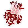
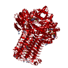
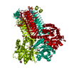
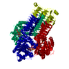
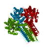
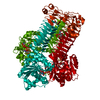
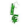
| Title | Crystal structure of GlmU from Mycobacterium tuberculosis in complex with uridine-diphosphate-N-acetylglucosamine | ||||||
 Components Components | Bifunctional protein glmU | ||||||
 Keywords Keywords | TRANSFERASE / Nucleotide-diphospho-sugar transferases/Single-stranded left-handed beta-helix / Acyltransferase / Cell shape / Cell wall biogenesis/degradation / Cytoplasm / Magnesium / Metal-binding / Multifunctional enzyme / Nucleotidyltransferase / Peptidoglycan synthesis | ||||||
| Function / homology |  Function and homology information Function and homology informationentry of bacterium into host cell / uridylyltransferase activity / adhesion of symbiont to host cell / glucosamine-1-phosphate N-acetyltransferase / glucosamine-1-phosphate N-acetyltransferase activity / UDP-N-acetylglucosamine diphosphorylase / UDP-N-acetylglucosamine diphosphorylase activity / UDP-N-acetylglucosamine biosynthetic process / lipid A biosynthetic process / peptidoglycan biosynthetic process ...entry of bacterium into host cell / uridylyltransferase activity / adhesion of symbiont to host cell / glucosamine-1-phosphate N-acetyltransferase / glucosamine-1-phosphate N-acetyltransferase activity / UDP-N-acetylglucosamine diphosphorylase / UDP-N-acetylglucosamine diphosphorylase activity / UDP-N-acetylglucosamine biosynthetic process / lipid A biosynthetic process / peptidoglycan biosynthetic process / cell wall organization / cell morphogenesis / regulation of cell shape / magnesium ion binding / membrane / cytosol / cytoplasm Similarity search - Function | ||||||
| Biological species |   Mycobacterium tuberculosis (bacteria) Mycobacterium tuberculosis (bacteria) | ||||||
| Method |  X-RAY DIFFRACTION / MOLECULAR REPLACEMENT / Resolution: 2.55 Å X-RAY DIFFRACTION / MOLECULAR REPLACEMENT / Resolution: 2.55 Å | ||||||
 Authors Authors | Zhang, Z. / Squire, C.J. / Baker, E.N. | ||||||
 Citation Citation | Journal: Acta Crystallogr.,Sect.D / Year: 2009 Title: Structure and function of GlmU from Mycobacterium tuberculosis. Authors: Zhang, Z. / Bulloch, E.M. / Bunker, R.D. / Baker, E.N. / Squire, C.J. | ||||||
| History |
|
- Structure visualization
| Structure viewer | Molecule: MolmilJmol/JSmol |
|---|
- Downloads & links
-Download
| PDBx/mmCIF format | 3d8v.cif.gz | 105.8 KB | Display | PDBx/mmCIF format |
|---|---|---|---|---|
| PDB format | pdb3d8v.ent.gz | 79.7 KB | Display | PDB format |
| PDBx/mmJSON format | 3d8v.json.gz | Tree view | PDBx/mmJSON format | |
| Others |  Other downloads Other downloads |
-Validation report
| Arichive directory | https://data.pdbj.org/pub/pdb/validation_reports/d8/3d8vftp://data.pdbj.org/pub/pdb/validation_reports/d8/3d8v | HTTPS FTP |
|---|
-Related structure data
| Related structure data |  2qkxC  3d98C  1hm9S C: citing same article ( S: Starting model for refinement |
|---|---|
| Similar structure data |

-Links
 PDBj
PDBj


- Assembly
| Deposited unit | 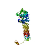
| ||||||||
|---|---|---|---|---|---|---|---|---|---|
| 1 | 
| ||||||||
| Unit cell |
|
-Components
| #1: Protein | Mass: 51637.727 Da / Num. of mol.: 1 Source method: isolated from a genetically manipulated source Source: (gene. exp.) Mycobacterium tuberculosis (bacteria) / Gene: glmU, Rv1018c, MT1046 / Plasmid: pDest17 / Production host: References: UniProt: P96382, UniProt: P9WMN3*PLUS, UDP-N-acetylglucosamine diphosphorylase, glucosamine-1-phosphate N-acetyltransferase |
|---|---|
| #2: Chemical | ChemComp-UD1 /   Mass: 607.354 Da / Num. of mol.: 1 / Source method: obtained synthetically / Formula: C17H27N3O17P2 Mass: 607.354 Da / Num. of mol.: 1 / Source method: obtained synthetically / Formula: C17H27N3O17P2 |
| #3: Water | ChemComp-HOH /  Mass: 18.015 Da / Num. of mol.: 147 / Source method: isolated from a natural source / Formula: H2O Mass: 18.015 Da / Num. of mol.: 147 / Source method: isolated from a natural source / Formula: H2O |
-Experimental details
-Experiment
| Experiment | Method: X-RAY DIFFRACTION / Number of used crystals: 1 |
|---|
- Sample preparation
| Crystal | Density Matthews: 4.4 Å3/Da / Density % sol: 72.04 % |
|---|---|
| Crystal grow | Temperature: 291 K / Method: vapor diffusion, sitting drop / pH: 7 Details: 5% PEG 8000, 0.1 M Cacodylate, 9% MPD, 12% Ethylene glycol, pH 7.0, VAPOR DIFFUSION, SITTING DROP, temperature 291K |
-Data collection
| Diffraction | Mean temperature: 113 K |
|---|---|
| Diffraction source | Source: ROTATING ANODE / Type: RIGAKU RU300 / Wavelength: 1.5417 Å |
| Detector | Type: MAR scanner 345 mm plate / Detector: IMAGE PLATE / Date: Jul 12, 2007 |
| Radiation | Monochromator: Osmic mirrors / Protocol: SINGLE WAVELENGTH / Monochromatic (M) / Laue (L): M / Scattering type: x-ray |
| Radiation wavelength | Wavelength: 1.5417 Å / Relative weight: 1 |
| Reflection | Resolution: 2.55→42.49 Å / Num. obs: 31984 / Observed criterion σ(I): 1 / Redundancy: 11.8 % / Biso Wilson estimate: 49.01 Å2 / Rmerge(I) obs: 0.11 / Net I/σ(I): 22.3 |
| Reflection shell | Resolution: 2.55→2.69 Å / Redundancy: 11.4 % / Rmerge(I) obs: 0.768 / Mean I/σ(I) obs: 2.3 / % possible all: 100 |
- Processing
| Software |
| ||||||||||||||||||||||||
|---|---|---|---|---|---|---|---|---|---|---|---|---|---|---|---|---|---|---|---|---|---|---|---|---|---|
| Refinement | Method to determine structure: MOLECULAR REPLACEMENT Starting model: PDB entry 1HM9 Resolution: 2.55→33.979 Å / SU ML: 0.32 / Isotropic thermal model: anisotropic / Phase error: 25.27 / Stereochemistry target values: Engh & Huber
| ||||||||||||||||||||||||
| Solvent computation | Shrinkage radii: 0.9 Å / VDW probe radii: 1.11 Å / Solvent model: FLAT BULK SOLVENT MODEL / Bsol: 49.341 Å2 / ksol: 0.329 e/Å3 | ||||||||||||||||||||||||
| Displacement parameters | Biso mean: 47 Å2
| ||||||||||||||||||||||||
| Refinement step | Cycle: LAST / Resolution: 2.55→33.979 Å
| ||||||||||||||||||||||||
| Refine LS restraints |
| ||||||||||||||||||||||||
| LS refinement shell | Resolution: 2.55→2.63 Å / Total num. of bins used: 11
|
