 Movie
Movie Controller

[English] 日本語
 Yorodumi
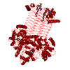
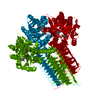
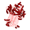
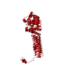
- PDB-4hcq: Crystal structure of GLMU from mycobacterium tuberculosis in comp... -
+ Open data

- Basic information
| Entry | Database: PDB / ID: 4hcq | |||||||||
|---|---|---|---|---|---|---|---|---|---|---|
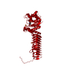
| Title | Crystal structure of GLMU from mycobacterium tuberculosis in complex with glucosamine-1-phosphate | |||||||||
 Components Components | Bifunctional protein GlmU | |||||||||
 Keywords Keywords | TRANSFERASE / ACETYLTRANSFERASE / BIFUNCTIONAL / PYROPHOSPHORYLASE / ROSSMANN-LIKE FOLD / LEFT-HANDED-BETA-HELIX / CELL SHAPE / CELL WALL BIOGENESIS/DEGRADATION / MAGNESIUM / METAL-BINDING / MULTIFUNCTIONAL ENZYME / NUCLEOTIDYLTRANSFERASE / PEPTIDOGLYCAN SYNTHESIS / ACYLTRANSFERASE | |||||||||
| Function / homology |  Function and homology information Function and homology informationentry of bacterium into host cell / uridylyltransferase activity / adhesion of symbiont to host cell / glucosamine-1-phosphate N-acetyltransferase / glucosamine-1-phosphate N-acetyltransferase activity / UDP-N-acetylglucosamine diphosphorylase / UDP-N-acetylglucosamine diphosphorylase activity / UDP-N-acetylglucosamine biosynthetic process / lipid A biosynthetic process / peptidoglycan biosynthetic process ...entry of bacterium into host cell / uridylyltransferase activity / adhesion of symbiont to host cell / glucosamine-1-phosphate N-acetyltransferase / glucosamine-1-phosphate N-acetyltransferase activity / UDP-N-acetylglucosamine diphosphorylase / UDP-N-acetylglucosamine diphosphorylase activity / UDP-N-acetylglucosamine biosynthetic process / lipid A biosynthetic process / peptidoglycan biosynthetic process / cell wall organization / cell morphogenesis / regulation of cell shape / magnesium ion binding / membrane / cytosol / cytoplasm Similarity search - Function | |||||||||
| Biological species |   Mycobacterium tuberculosis (bacteria) Mycobacterium tuberculosis (bacteria) | |||||||||
| Method |  X-RAY DIFFRACTION / MOLECULAR REPLACEMENT / Resolution: 2.6 Å X-RAY DIFFRACTION / MOLECULAR REPLACEMENT / Resolution: 2.6 Å | |||||||||
 Authors Authors | Jagtap, P.K.A. / Verma, S.K. / Vithani, N. | |||||||||
 Citation Citation | Journal: J.Mol.Biol. / Year: 2013 Title: Crystal structures identify an atypical two-metal-ion mechanism for uridyltransfer in GlmU: its significance to sugar nucleotidyl transferases Authors: Jagtap, P.K.A. / Verma, S.K. / Vithani, N. / Bais, V.S. / Prakash, B. | |||||||||
| History |
|
- Structure visualization
| Structure viewer | Molecule: MolmilJmol/JSmol |
|---|
- Downloads & links
-Download
| PDBx/mmCIF format | 4hcq.cif.gz | 101.6 KB | Display | PDBx/mmCIF format |
|---|---|---|---|---|
| PDB format | pdb4hcq.ent.gz | 75.5 KB | Display | PDB format |
| PDBx/mmJSON format | 4hcq.json.gz | Tree view | PDBx/mmJSON format | |
| Others |  Other downloads Other downloads |
-Validation report
| Arichive directory | https://data.pdbj.org/pub/pdb/validation_reports/hc/4hcqftp://data.pdbj.org/pub/pdb/validation_reports/hc/4hcq | HTTPS FTP |
|---|
-Related structure data
| Related structure data | 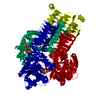 4g87C 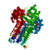 3dj4S S: Starting model for refinement C: citing same article ( |
|---|---|
| Similar structure data |

-Links
 PDBj
PDBj


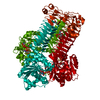
- Assembly
| Deposited unit | 
| ||||||||
|---|---|---|---|---|---|---|---|---|---|
| 1 | 
| ||||||||
| Unit cell |
|
-Components
| #1: Protein | Mass: 52466.602 Da / Num. of mol.: 1 Source method: isolated from a genetically manipulated source Source: (gene. exp.) Mycobacterium tuberculosis (bacteria) / Strain: H37RV / Gene: glmU, Rv1018c / Plasmid: PQEII / Production host: References: UniProt: P96382, UniProt: P9WMN3*PLUS, UDP-N-acetylglucosamine diphosphorylase, glucosamine-1-phosphate N-acetyltransferase | ||||||||
|---|---|---|---|---|---|---|---|---|---|
| #2: Sugar |   Type: D-saccharide / Mass: 301.188 Da / Num. of mol.: 2 Type: D-saccharide / Mass: 301.188 Da / Num. of mol.: 2Source method: isolated from a genetically manipulated source Formula: C8H16NO9P #3: Chemical | ChemComp-CO / |   Mass: 58.933 Da / Num. of mol.: 1 / Source method: obtained synthetically / Formula: Co Mass: 58.933 Da / Num. of mol.: 1 / Source method: obtained synthetically / Formula: Co#4: Chemical |   Mass: 24.305 Da / Num. of mol.: 2 / Source method: obtained synthetically / Formula: Mg Mass: 24.305 Da / Num. of mol.: 2 / Source method: obtained synthetically / Formula: Mg#5: Water | ChemComp-HOH / |  Mass: 18.015 Da / Num. of mol.: 87 / Source method: isolated from a natural source / Formula: H2O Mass: 18.015 Da / Num. of mol.: 87 / Source method: isolated from a natural source / Formula: H2OHas protein modification | N | |
-Experimental details
-Experiment
| Experiment | Method: X-RAY DIFFRACTION / Number of used crystals: 1 |
|---|
- Sample preparation
| Crystal | Density Matthews: 3.19 Å3/Da / Density % sol: 61.43 % |
|---|---|
| Crystal grow | Temperature: 277 K / Method: vapor diffusion / pH: 7.5 Details: 8% PEG 8000, 150MM NACL, 5% GLYCEROL, 1,3-BUTANEDIOL, AMPPNP, MGCL2, 0.1M HEPES, COCL2, DTT, pH 7.5, VAPOR DIFFUSION, temperature 277K |
-Data collection
| Diffraction | Mean temperature: 100 K |
|---|---|
| Diffraction source | Source: ROTATING ANODE / Type: RIGAKU MICROMAX-007 HF / Wavelength: 1.54 |
| Detector | Type: MAR scanner 345 mm plate / Detector: IMAGE PLATE / Date: Feb 9, 2009 / Details: MIRRORS |
| Radiation | Protocol: SINGLE WAVELENGTH / Monochromatic (M) / Laue (L): M / Scattering type: x-ray |
| Radiation wavelength | Wavelength: 1.54 Å / Relative weight: 1 |
| Reflection | Resolution: 2.6→19.932 Å / Num. obs: 19893 / % possible obs: 99.4 % / Observed criterion σ(I): -3 / Biso Wilson estimate: 42.49 Å2 / Rmerge(I) obs: 0.088 / Net I/σ(I): 20.1 |
| Reflection shell | Resolution: 2.6→2.67 Å / Rmerge(I) obs: 0.602 / Mean I/σ(I) obs: 2.86 / % possible all: 94.9 |
- Processing
| Software |
| ||||||||||||||||||||||||||||||||||||||||||||||||||||||||||||||||||||||||||||||||||||||||||||||||||||||||||||||||||||||||||||||||||||||||||||||||||||||||||||||||||||||||||
|---|---|---|---|---|---|---|---|---|---|---|---|---|---|---|---|---|---|---|---|---|---|---|---|---|---|---|---|---|---|---|---|---|---|---|---|---|---|---|---|---|---|---|---|---|---|---|---|---|---|---|---|---|---|---|---|---|---|---|---|---|---|---|---|---|---|---|---|---|---|---|---|---|---|---|---|---|---|---|---|---|---|---|---|---|---|---|---|---|---|---|---|---|---|---|---|---|---|---|---|---|---|---|---|---|---|---|---|---|---|---|---|---|---|---|---|---|---|---|---|---|---|---|---|---|---|---|---|---|---|---|---|---|---|---|---|---|---|---|---|---|---|---|---|---|---|---|---|---|---|---|---|---|---|---|---|---|---|---|---|---|---|---|---|---|---|---|---|---|---|---|---|
| Refinement | Method to determine structure: MOLECULAR REPLACEMENT Starting model: 3DJ4 Resolution: 2.6→19.93 Å / Cor.coef. Fo:Fc: 0.937 / Cor.coef. Fo:Fc free: 0.886 / SU B: 9.309 / SU ML: 0.2 / Cross valid method: THROUGHOUT / σ(F): 0 / ESU R Free: 0.299 / Stereochemistry target values: MAXIMUM LIKELIHOOD Details: HYDROGENS HAVE BEEN ADDED IN THE RIDING POSITIONS U VALUES: REFINED INDIVIDUALLY
| ||||||||||||||||||||||||||||||||||||||||||||||||||||||||||||||||||||||||||||||||||||||||||||||||||||||||||||||||||||||||||||||||||||||||||||||||||||||||||||||||||||||||||
| Solvent computation | Ion probe radii: 0.8 Å / Shrinkage radii: 0.8 Å / VDW probe radii: 1.4 Å / Solvent model: MASK | ||||||||||||||||||||||||||||||||||||||||||||||||||||||||||||||||||||||||||||||||||||||||||||||||||||||||||||||||||||||||||||||||||||||||||||||||||||||||||||||||||||||||||
| Displacement parameters | Biso mean: 33.01 Å2
| ||||||||||||||||||||||||||||||||||||||||||||||||||||||||||||||||||||||||||||||||||||||||||||||||||||||||||||||||||||||||||||||||||||||||||||||||||||||||||||||||||||||||||
| Refinement step | Cycle: LAST / Resolution: 2.6→19.93 Å
| ||||||||||||||||||||||||||||||||||||||||||||||||||||||||||||||||||||||||||||||||||||||||||||||||||||||||||||||||||||||||||||||||||||||||||||||||||||||||||||||||||||||||||
| Refine LS restraints |
| ||||||||||||||||||||||||||||||||||||||||||||||||||||||||||||||||||||||||||||||||||||||||||||||||||||||||||||||||||||||||||||||||||||||||||||||||||||||||||||||||||||||||||
| LS refinement shell | Resolution: 2.6→2.67 Å / Total num. of bins used: 20
|
